 Movie
Movie Controller

[English] 日本語
 Yorodumi
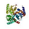
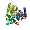
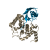
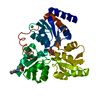
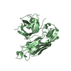
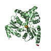
- PDB-5lqm: Structure of quinolinate synthase Y21F mutant in complex with citrate -
+ Open data

- Basic information
| Entry | Database: PDB / ID: 5lqm | ||||||
|---|---|---|---|---|---|---|---|
| Title | Structure of quinolinate synthase Y21F mutant in complex with citrate | ||||||
 Components Components | Quinolinate synthase A | ||||||
 Keywords Keywords |  TRANSFERASE / NAD BIOSYNTHESIS / IRON SULFUR CLUSTER TRANSFERASE / NAD BIOSYNTHESIS / IRON SULFUR CLUSTER | ||||||
| Function / homology |  Function and homology informationquinolinate synthase / quinolinate synthetase A activity / 'de novo' NAD biosynthetic process from aspartate / 4 iron, 4 sulfur cluster binding / metal ion binding / cytosol Function and homology informationquinolinate synthase / quinolinate synthetase A activity / 'de novo' NAD biosynthetic process from aspartate / 4 iron, 4 sulfur cluster binding / metal ion binding / cytosolSimilarity search - Function | ||||||
| Biological species |   Thermotoga maritima (bacteria) Thermotoga maritima (bacteria) | ||||||
| Method | X-RAY DIFFRACTION / SYNCHROTRON / MOLECULAR REPLACEMENT / Resolution: 1.62 Å | ||||||
 Authors Authors | Volbeda, A. / Fontecilla-Camps, J.C. | ||||||
| Funding support |  France, 1items France, 1items
| ||||||
 Citation Citation | Journal: J.Am.Chem.Soc. / Year: 2016 Title: Crystal Structures of Quinolinate Synthase in Complex with a Substrate Analogue, the Condensation Intermediate, and Substrate-Derived Product. Authors: Volbeda, A. / Darnault, C. / Renoux, O. / Reichmann, D. / Amara, P. / Ollagnier de Choudens, S. / Fontecilla-Camps, J.C. #1: Journal: J. Am. Chem. Soc. / Year: 2014Title: The crystal structure of Fe4S4 quinolinate synthase unravels an enzymatic dehydration mechanism that uses tyrosine and a hydrolase-type triad. Authors: Cherrier, M.V. / Chan, A. / Darnault, C. / Reichmann, D. / Amara, P. / Ollagnier de Choudens, S. / Fontecilla-Camps, J.C. | ||||||
| History |
|
- Structure visualization
| Structure viewer | Molecule: MolmilJmol/JSmol |
|---|
- Downloads & links
-Download
| PDBx/mmCIF format | 5lqm.cif.gz | 150.9 KB | Display | PDBx/mmCIF format |
|---|---|---|---|---|
| PDB format | pdb5lqm.ent.gz | 117.1 KB | Display | PDB format |
| PDBx/mmJSON format | 5lqm.json.gz | Tree view | PDBx/mmJSON format | |
| Others |  Other downloads Other downloads |
-Validation report
| Arichive directory | https://data.pdbj.org/pub/pdb/validation_reports/lq/5lqmftp://data.pdbj.org/pub/pdb/validation_reports/lq/5lqm | HTTPS FTP |
|---|
-Related structure data
| Related structure data |  5f33C 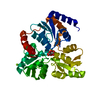 5f35C  5f3dC  5lqsC  4p3xS S: Starting model for refinement C: citing same article ( |
|---|---|
| Similar structure data |


-Links
 PDBj
PDBj


- Assembly
| Deposited unit | 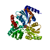
| ||||||||
|---|---|---|---|---|---|---|---|---|---|
| 1 |
| ||||||||
| Unit cell |
|
-Components
| #1: Protein | Mass: 34640.598 Da / Num. of mol.: 1 Source method: isolated from a genetically manipulated source Details: Specific_mutation = "Y21F " Specific_mutation = "K219R " Source: (gene. exp.) Thermotoga maritima (strain ATCC 43589 / MSB8 / DSM 3109 / JCM 10099) (bacteria)Strain: ATCC 43589 / MSB8 / DSM 3109 / JCM 10099 / Gene: nadA, TM_1644 / Plasmid: PT7 Production host: Escherichia coli 'BL21-Gold(DE3)pLysS AG' (bacteria)References: UniProt: Q9X1X7, quinolinate synthase |
|---|---|
| #2: Chemical | ChemComp-SF4 / Iron–sulfur cluster  Mass: 351.640 Da / Num. of mol.: 1 / Source method: obtained synthetically / Formula: Fe4S4 Mass: 351.640 Da / Num. of mol.: 1 / Source method: obtained synthetically / Formula: Fe4S4 |
| #3: Chemical | ChemComp-FLC / Citric acid  Mass: 189.100 Da / Num. of mol.: 1 / Source method: obtained synthetically / Formula: C6H5O7 Mass: 189.100 Da / Num. of mol.: 1 / Source method: obtained synthetically / Formula: C6H5O7 |
| #4: Water | ChemComp-HOH / Water Mass: 18.015 Da / Num. of mol.: 282 / Source method: isolated from a natural source / Formula: H2O Mass: 18.015 Da / Num. of mol.: 282 / Source method: isolated from a natural source / Formula: H2O |
-Experimental details
-Experiment
| Experiment | Method: X-RAY DIFFRACTION / Number of used crystals: 1 |
|---|
- Sample preparation
| Crystal | Density Matthews: 2.18 Å3/Da / Density % sol: 43.7 % |
|---|---|
| Crystal grow | Temperature: 298 K / Method: vapor diffusion / Details: PEG3350, sodium citrate, KCl, anaerobic |
-Data collection
| Diffraction | Mean temperature: 100 K |
|---|---|
| Diffraction source | Source: SYNCHROTRON / Site: ESRF / Beamline: ID30B / Wavelength: 0.96862 Å |
| Detector | Type: DECTRIS PILATUS 6M-F / Detector: PIXEL / Date: Oct 31, 2015 |
| Radiation | Protocol: SINGLE WAVELENGTH / Monochromatic (M) / Laue (L): M / Scattering type: x-ray |
| Radiation wavelength | Wavelength: 0.96862 Å / Relative weight: 1 |
| Reflection | Resolution: 1.62→37.53 Å / Num. obs: 34681 / % possible obs: 91 % / Redundancy: 2.5 % / CC1/2: 0.998 / Rmerge(I) obs: 0.044 / Net I/σ(I): 12 |
| Reflection shell | Resolution: 1.62→1.68 Å / Redundancy: 2.4 % / Rmerge(I) obs: 0.454 / Mean I/σ(I) obs: 1.8 / CC1/2: 0.49 / % possible all: 62 |
- Processing
| Software |
| |||||||||||||||||||||||||||||||||||||||||||||||||||||||||||||||||||||||||||||||||||||
|---|---|---|---|---|---|---|---|---|---|---|---|---|---|---|---|---|---|---|---|---|---|---|---|---|---|---|---|---|---|---|---|---|---|---|---|---|---|---|---|---|---|---|---|---|---|---|---|---|---|---|---|---|---|---|---|---|---|---|---|---|---|---|---|---|---|---|---|---|---|---|---|---|---|---|---|---|---|---|---|---|---|---|---|---|---|---|
| Refinement | Method to determine structure: MOLECULAR REPLACEMENT Starting model: 4P3X Resolution: 1.62→37.53 Å / Cor.coef. Fo:Fc: 0.975 / Cor.coef. Fo:Fc free: 0.957 / SU B: 5.674 / SU ML: 0.081 / Cross valid method: THROUGHOUT / ESU R: 0.15 / ESU R Free: 0.099 / Details: HYDROGENS HAVE BEEN ADDED IN THE RIDING POSITIONS
| |||||||||||||||||||||||||||||||||||||||||||||||||||||||||||||||||||||||||||||||||||||
| Solvent computation | Ion probe radii: 0.8 Å / Shrinkage radii: 0.8 Å / VDW probe radii: 1.2 Å | |||||||||||||||||||||||||||||||||||||||||||||||||||||||||||||||||||||||||||||||||||||
| Displacement parameters | Biso mean: 23.029 Å2
| |||||||||||||||||||||||||||||||||||||||||||||||||||||||||||||||||||||||||||||||||||||
| Refinement step | Cycle: 1 / Resolution: 1.62→37.53 Å
| |||||||||||||||||||||||||||||||||||||||||||||||||||||||||||||||||||||||||||||||||||||
| Refine LS restraints |
| |||||||||||||||||||||||||||||||||||||||||||||||||||||||||||||||||||||||||||||||||||||
| LS refinement shell | Resolution: 1.62→1.662 Å / Total num. of bins used: 20
|
