 Movie
Movie Controller

[English] 日本語
 Yorodumi
- PDB-5lqj: Crystal Structure of COMT in complex with 3-cyclopropyl-5-methyl-... -
+ Open data

- Basic information
| Entry | Database: PDB / ID: 5lqj | ||||||
|---|---|---|---|---|---|---|---|
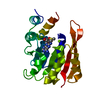
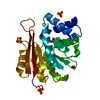
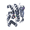
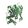
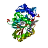
| Title | Crystal Structure of COMT in complex with 3-cyclopropyl-5-methyl-4-phenyl-1,2,4-triazole | ||||||
 Components Components | Catechol O-methyltransferase Catechol-O-methyltransferase Catechol-O-methyltransferase | ||||||
 Keywords Keywords | TRANSFERASE / METHYLTRANSFERASE / NEUROTRANSMITTER DEGRADATION / CATECHOL | ||||||
| Function / homology |  Function and homology information Function and homology informationEnzymatic degradation of dopamine by COMT / Enzymatic degradation of Dopamine by monoamine oxidase / positive regulation of homocysteine metabolic process / Methylation / norepinephrine secretion / response to dopamine / mastication / catecholamine catabolic process / catechol-containing compound metabolic process / catechol O-methyltransferase activity ...Enzymatic degradation of dopamine by COMT / Enzymatic degradation of Dopamine by monoamine oxidase / positive regulation of homocysteine metabolic process / Methylation / norepinephrine secretion / response to dopamine / mastication / catecholamine catabolic process / catechol-containing compound metabolic process / catechol O-methyltransferase activity / renal sodium excretion / : / : / S-adenosylhomocysteine metabolic process / catechol O-methyltransferase / developmental process / renal filtration / renin secretion into blood stream / dopamine secretion / renal albumin absorption / negative regulation of dopamine metabolic process / response to salt / habituation / artery development / catecholamine metabolic process / S-adenosylmethionine metabolic process / short-term memory / cerebellar cortex morphogenesis / cellular response to phosphate starvation / dopamine catabolic process / norepinephrine metabolic process / glomerulus development / fear response / multicellular organismal reproductive process / synaptic transmission, dopaminergic / response to angiotensin / cellular response to cocaine / estrogen metabolic process / response to food / exploration behavior / cholesterol efflux / response to temperature stimulus / response to pain / response to corticosterone / dopamine metabolic process / glycogen metabolic process / prostaglandin metabolic process / startle response / detection of temperature stimulus involved in sensory perception of pain / : / behavioral fear response / multicellular organismal response to stress / response to amphetamine / : / kidney development / learning / response to cytokine / female pregnancy / negative regulation of smooth muscle cell proliferation / visual learning / multicellular organism growth / response to organic cyclic compound / response to toxic substance / memory / cognition / regulation of blood pressure / response to wounding / response to estrogen / gene expression / cell body / methylation / postsynapse / postsynaptic membrane / response to oxidative stress / vesicle / response to lipopolysaccharide / dendritic spine / learning or memory / response to hypoxia / response to xenobiotic stimulus / axon / glutamatergic synapse / dendrite / magnesium ion binding / membrane / cytosolSimilarity search - Function | ||||||
| Biological species |  Rattus norvegicus (Norway rat) Rattus norvegicus (Norway rat) | ||||||
| Method | X-RAY DIFFRACTION / SYNCHROTRON / MOLECULAR REPLACEMENT / Resolution: 2.41 Å | ||||||
 Authors Authors | Ehler, A. / Lerner, C. / Rudolph, M.G. | ||||||
 Citation Citation | Journal: To be published Title: Crystal Structure of COMT in complex with 3-cyclopropyl-5-methyl-4-phenyl-1,2,4-triazole Authors: Lerner, C. / Rudolph, M.G. | ||||||
| History |
|
- Structure visualization
| Structure viewer | Molecule: MolmilJmol/JSmol |
|---|
- Downloads & links
-Download
| PDBx/mmCIF format | 5lqj.cif.gz | 350 KB | Display | PDBx/mmCIF format |
|---|---|---|---|---|
| PDB format | pdb5lqj.ent.gz | 287.4 KB | Display | PDB format |
| PDBx/mmJSON format | 5lqj.json.gz | Tree view | PDBx/mmJSON format | |
| Others |  Other downloads Other downloads |
-Validation report
| Arichive directory | https://data.pdbj.org/pub/pdb/validation_reports/lq/5lqjftp://data.pdbj.org/pub/pdb/validation_reports/lq/5lqj | HTTPS FTP |
|---|
-Related structure data
| Similar structure data |
|---|


-Links
 PDBj
PDBj- Assembly
| Deposited unit | 
| ||||||||
|---|---|---|---|---|---|---|---|---|---|
| 1 | 
| ||||||||
| 2 | 
| ||||||||
| 3 | 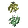
| ||||||||
| 4 | 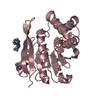
| ||||||||
| Unit cell |
|
-Components
-Protein , 1 types, 4 molecules ABCD
| #1: Protein | Catechol-O-methyltransferase Mass: 24694.332 Da / Num. of mol.: 4 / Fragment: SOLUBLE FORM, RESIDUES 44-264 Source method: isolated from a genetically manipulated source Source: (gene. exp.) Rattus norvegicus (Norway rat) / Gene: Comt / Production host:  Escherichia coli BL21(DE3) (bacteria) / References: UniProt: P22734, catechol O-methyltransferase Escherichia coli BL21(DE3) (bacteria) / References: UniProt: P22734, catechol O-methyltransferase |
|---|
-Non-polymers , 5 types, 136 molecules 


| #2: Chemical |  Mass: 22.990 Da / Num. of mol.: 3 / Source method: obtained synthetically / Formula: Na Mass: 22.990 Da / Num. of mol.: 3 / Source method: obtained synthetically / Formula: Na#3: Chemical | ChemComp-72N /  Mass: 199.252 Da / Num. of mol.: 5 / Source method: obtained synthetically / Formula: C12H13N3 Mass: 199.252 Da / Num. of mol.: 5 / Source method: obtained synthetically / Formula: C12H13N3#4: Chemical | ChemComp-CL / | Chloride Mass: 35.453 Da / Num. of mol.: 1 / Source method: obtained synthetically / Formula: Cl Mass: 35.453 Da / Num. of mol.: 1 / Source method: obtained synthetically / Formula: Cl#5: Chemical | ChemComp-K / |  Mass: 39.098 Da / Num. of mol.: 1 / Source method: obtained synthetically / Formula: K Mass: 39.098 Da / Num. of mol.: 1 / Source method: obtained synthetically / Formula: K#6: Water | ChemComp-HOH / | WaterMass: 18.015 Da / Num. of mol.: 126 / Source method: isolated from a natural source / Formula: H2O |
|---|
-Experimental details
-Experiment
| Experiment | Method: X-RAY DIFFRACTION / Number of used crystals: 1 |
|---|
- Sample preparation
| Crystal | Density Matthews: 2.52 Å3/Da / Density % sol: 51.28 % |
|---|---|
| Crystal grow | Temperature: 295 K / Method: vapor diffusion, sitting drop / pH: 9 / Details: AMMONIUM SULPHATE, CHES, PH 9 |
-Data collection
| Diffraction | Mean temperature: 100 K | ||||||||||||||||||||||||||||||||||||||||||||||||||||||||||||||||||||||||||||||||||||||||||||||||||||||||||||||||||||||||||||||
|---|---|---|---|---|---|---|---|---|---|---|---|---|---|---|---|---|---|---|---|---|---|---|---|---|---|---|---|---|---|---|---|---|---|---|---|---|---|---|---|---|---|---|---|---|---|---|---|---|---|---|---|---|---|---|---|---|---|---|---|---|---|---|---|---|---|---|---|---|---|---|---|---|---|---|---|---|---|---|---|---|---|---|---|---|---|---|---|---|---|---|---|---|---|---|---|---|---|---|---|---|---|---|---|---|---|---|---|---|---|---|---|---|---|---|---|---|---|---|---|---|---|---|---|---|---|---|---|
| Diffraction source | Source: SYNCHROTRON / Site: SLS  / Beamline: X10SA / Wavelength: 1 Å / Beamline: X10SA / Wavelength: 1 Å | ||||||||||||||||||||||||||||||||||||||||||||||||||||||||||||||||||||||||||||||||||||||||||||||||||||||||||||||||||||||||||||||
| Detector | Type: PSI PILATUS 6M / Detector: PIXEL / Date: May 22, 2010 | ||||||||||||||||||||||||||||||||||||||||||||||||||||||||||||||||||||||||||||||||||||||||||||||||||||||||||||||||||||||||||||||
| Radiation | Protocol: SINGLE WAVELENGTH / Monochromatic (M) / Laue (L): M / Scattering type: x-ray | ||||||||||||||||||||||||||||||||||||||||||||||||||||||||||||||||||||||||||||||||||||||||||||||||||||||||||||||||||||||||||||||
| Radiation wavelength | Wavelength: 1 Å / Relative weight: 1 | ||||||||||||||||||||||||||||||||||||||||||||||||||||||||||||||||||||||||||||||||||||||||||||||||||||||||||||||||||||||||||||||
| Reflection | Resolution: 2.41→120.3 Å / Num. obs: 39635 / % possible obs: 99.9 % / Observed criterion σ(I): -3 / Redundancy: 13.3 % / Biso Wilson estimate: 57.81 Å2 / CC1/2: 0.998 / Rmerge(I) obs: 0.217 / Net I/σ(I): 11.21 | ||||||||||||||||||||||||||||||||||||||||||||||||||||||||||||||||||||||||||||||||||||||||||||||||||||||||||||||||||||||||||||||
| Reflection shell |
|
- Processing
| Software |
| |||||||||||||||||||||||||||||||||||||||||||||||||||||||||||||||||||||||||||||||||||||||||||||||||||||||||||||||||||||||||||||||||||||||||||||||||||||||||||||||||||||||||||||||||||||||||||||||||||||||||||||||||||||||||||||||||||||||||||||||||||||||||||||||||||||||||||||||||||||||||||||||||||||||||||||||||||||||||||||||||||||||||||||||||||||||||||||||||||||||||||||||||||||||||||||||||||||||||||||||||||||||||||||||||||||||||||||||||||||||||||||||||||||||||||||||||||||||||||
|---|---|---|---|---|---|---|---|---|---|---|---|---|---|---|---|---|---|---|---|---|---|---|---|---|---|---|---|---|---|---|---|---|---|---|---|---|---|---|---|---|---|---|---|---|---|---|---|---|---|---|---|---|---|---|---|---|---|---|---|---|---|---|---|---|---|---|---|---|---|---|---|---|---|---|---|---|---|---|---|---|---|---|---|---|---|---|---|---|---|---|---|---|---|---|---|---|---|---|---|---|---|---|---|---|---|---|---|---|---|---|---|---|---|---|---|---|---|---|---|---|---|---|---|---|---|---|---|---|---|---|---|---|---|---|---|---|---|---|---|---|---|---|---|---|---|---|---|---|---|---|---|---|---|---|---|---|---|---|---|---|---|---|---|---|---|---|---|---|---|---|---|---|---|---|---|---|---|---|---|---|---|---|---|---|---|---|---|---|---|---|---|---|---|---|---|---|---|---|---|---|---|---|---|---|---|---|---|---|---|---|---|---|---|---|---|---|---|---|---|---|---|---|---|---|---|---|---|---|---|---|---|---|---|---|---|---|---|---|---|---|---|---|---|---|---|---|---|---|---|---|---|---|---|---|---|---|---|---|---|---|---|---|---|---|---|---|---|---|---|---|---|---|---|---|---|---|---|---|---|---|---|---|---|---|---|---|---|---|---|---|---|---|---|---|---|---|---|---|---|---|---|---|---|---|---|---|---|---|---|---|---|---|---|---|---|---|---|---|---|---|---|---|---|---|---|---|---|---|---|---|---|---|---|---|---|---|---|---|---|---|---|---|---|---|---|---|---|---|---|---|---|---|---|---|---|---|---|---|---|---|---|---|---|---|---|---|---|---|---|---|---|---|---|---|---|---|---|---|---|---|---|---|---|---|---|---|---|---|---|---|---|---|---|---|---|---|---|---|---|---|---|---|---|---|---|---|---|---|---|---|---|---|---|---|---|---|---|---|---|---|---|---|---|---|---|---|---|---|---|---|---|---|---|---|---|---|---|---|---|---|---|---|---|---|---|---|---|---|---|---|---|---|---|---|---|---|---|---|---|---|---|---|---|---|---|---|---|---|---|---|---|---|---|---|---|---|
| Refinement | Method to determine structure: MOLECULAR REPLACEMENT Starting model: inhouse model Resolution: 2.41→47.201 Å / SU ML: 0.41 / Cross valid method: THROUGHOUT / σ(F): 1.33 / Phase error: 26.83 Details: ligand does not bind at the active site but acts as an additive for crystallization. Two of the four molecules in the a.u. have their C-terminal beta-strand domain-swapped.
| |||||||||||||||||||||||||||||||||||||||||||||||||||||||||||||||||||||||||||||||||||||||||||||||||||||||||||||||||||||||||||||||||||||||||||||||||||||||||||||||||||||||||||||||||||||||||||||||||||||||||||||||||||||||||||||||||||||||||||||||||||||||||||||||||||||||||||||||||||||||||||||||||||||||||||||||||||||||||||||||||||||||||||||||||||||||||||||||||||||||||||||||||||||||||||||||||||||||||||||||||||||||||||||||||||||||||||||||||||||||||||||||||||||||||||||||||||||||||||
| Solvent computation | Shrinkage radii: 0.9 Å / VDW probe radii: 1.11 Å | |||||||||||||||||||||||||||||||||||||||||||||||||||||||||||||||||||||||||||||||||||||||||||||||||||||||||||||||||||||||||||||||||||||||||||||||||||||||||||||||||||||||||||||||||||||||||||||||||||||||||||||||||||||||||||||||||||||||||||||||||||||||||||||||||||||||||||||||||||||||||||||||||||||||||||||||||||||||||||||||||||||||||||||||||||||||||||||||||||||||||||||||||||||||||||||||||||||||||||||||||||||||||||||||||||||||||||||||||||||||||||||||||||||||||||||||||||||||||||
| Displacement parameters | Biso max: 161.54 Å2 / Biso mean: 56.9636 Å2 / Biso min: 26.12 Å2 | |||||||||||||||||||||||||||||||||||||||||||||||||||||||||||||||||||||||||||||||||||||||||||||||||||||||||||||||||||||||||||||||||||||||||||||||||||||||||||||||||||||||||||||||||||||||||||||||||||||||||||||||||||||||||||||||||||||||||||||||||||||||||||||||||||||||||||||||||||||||||||||||||||||||||||||||||||||||||||||||||||||||||||||||||||||||||||||||||||||||||||||||||||||||||||||||||||||||||||||||||||||||||||||||||||||||||||||||||||||||||||||||||||||||||||||||||||||||||||
| Refinement step | Cycle: final / Resolution: 2.41→47.201 Å
| |||||||||||||||||||||||||||||||||||||||||||||||||||||||||||||||||||||||||||||||||||||||||||||||||||||||||||||||||||||||||||||||||||||||||||||||||||||||||||||||||||||||||||||||||||||||||||||||||||||||||||||||||||||||||||||||||||||||||||||||||||||||||||||||||||||||||||||||||||||||||||||||||||||||||||||||||||||||||||||||||||||||||||||||||||||||||||||||||||||||||||||||||||||||||||||||||||||||||||||||||||||||||||||||||||||||||||||||||||||||||||||||||||||||||||||||||||||||||||
| Refinement TLS params. | Method: refined / Refine-ID: X-RAY DIFFRACTION
| |||||||||||||||||||||||||||||||||||||||||||||||||||||||||||||||||||||||||||||||||||||||||||||||||||||||||||||||||||||||||||||||||||||||||||||||||||||||||||||||||||||||||||||||||||||||||||||||||||||||||||||||||||||||||||||||||||||||||||||||||||||||||||||||||||||||||||||||||||||||||||||||||||||||||||||||||||||||||||||||||||||||||||||||||||||||||||||||||||||||||||||||||||||||||||||||||||||||||||||||||||||||||||||||||||||||||||||||||||||||||||||||||||||||||||||||||||||||||||
| Refinement TLS group |
|