 Movie
Movie Controller


+ Open data

- Basic information
| Entry | Database: PDB / ID: 5ljj | ||||||||||||
|---|---|---|---|---|---|---|---|---|---|---|---|---|---|
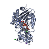
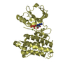
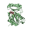
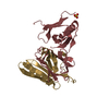
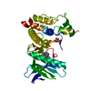
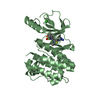
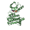
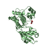
| Title | Crystal structure of human Mps1 (TTK) in complex with Reversine | ||||||||||||
 Components Components | Dual specificity protein kinase TTK | ||||||||||||
 Keywords Keywords |  TRANSFERASE / Mps1 / Reversine / TTK / kinase / mitosis checkpoint TRANSFERASE / Mps1 / Reversine / TTK / kinase / mitosis checkpoint | ||||||||||||
| Function / homology |  Function and homology information Function and homology informationprotein localization to meiotic spindle midzone / meiotic spindle assembly checkpoint signaling / kinetochore binding / female meiosis chromosome segregation / protein localization to kinetochore / dual-specificity kinase / spindle organization / mitotic spindle assembly checkpoint signaling / protein serine/threonine/tyrosine kinase activity / mitotic spindle organization ...protein localization to meiotic spindle midzone / meiotic spindle assembly checkpoint signaling / kinetochore binding / female meiosis chromosome segregation / protein localization to kinetochore / dual-specificity kinase / spindle organization / mitotic spindle assembly checkpoint signaling / protein serine/threonine/tyrosine kinase activity / mitotic spindle organization / chromosome segregation / kinetochore / spindle / protein tyrosine kinase activity / phosphorylation / protein serine kinase activity / protein serine/threonine kinase activity / positive regulation of cell population proliferation / ATP binding / membrane / identical protein binding / nucleus / cytoplasmSimilarity search - Function | ||||||||||||
| Biological species |  Homo sapiens (human) Homo sapiens (human) | ||||||||||||
| Method | X-RAY DIFFRACTION / SYNCHROTRON / MOLECULAR REPLACEMENT / molecular replacement / Resolution: 3 Å | ||||||||||||
 Authors Authors | Hiruma, Y. / Joosten, R.P. / Perrakis, A. | ||||||||||||
| Funding support |  Netherlands, 3items Netherlands, 3items
| ||||||||||||
 Citation Citation | Journal: Proteins / Year: 2016 Title: Structural basis of reversine selectivity in inhibiting Mps1 more potently than aurora B kinase. Authors: Hiruma, Y. / Koch, A. / Dharadhar, S. / Joosten, R.P. / Perrakis, A. | ||||||||||||
| History |
|
- Structure visualization
| Structure viewer | Molecule: MolmilJmol/JSmol |
|---|
- Downloads & links
-Download
| PDBx/mmCIF format | 5ljj.cif.gz | 127.1 KB | Display | PDBx/mmCIF format |
|---|---|---|---|---|
| PDB format | pdb5ljj.ent.gz | 97.4 KB | Display | PDB format |
| PDBx/mmJSON format | 5ljj.json.gz | Tree view | PDBx/mmJSON format | |
| Others |  Other downloads Other downloads |
-Validation report
| Arichive directory | https://data.pdbj.org/pub/pdb/validation_reports/lj/5ljjftp://data.pdbj.org/pub/pdb/validation_reports/lj/5ljj | HTTPS FTP |
|---|
-Related structure data
| Related structure data |  3hmnS S: Starting model for refinement |
|---|---|
| Similar structure data |


-Links
 PDBj
PDBj- Assembly
| Deposited unit | 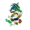
| ||||||||
|---|---|---|---|---|---|---|---|---|---|
| 1 |
| ||||||||
| Unit cell |
|
-Components
| #1: Protein | Mass: 36175.289 Da / Num. of mol.: 1 / Mutation: C604Y Source method: isolated from a genetically manipulated source Source: (gene. exp.) Homo sapiens (human) / Gene: TTK, MPS1, MPS1L1 / Production host:  Escherichia coli BL21(DE3) (bacteria) / References: UniProt: P33981, dual-specificity kinase Escherichia coli BL21(DE3) (bacteria) / References: UniProt: P33981, dual-specificity kinase |
|---|---|
| #2: Chemical | ChemComp-AD5 / Reversine  Mass: 393.485 Da / Num. of mol.: 1 / Source method: obtained synthetically / Formula: C21H27N7O / Comment: antagonist*YM Mass: 393.485 Da / Num. of mol.: 1 / Source method: obtained synthetically / Formula: C21H27N7O / Comment: antagonist*YM |
| #3: Chemical | ChemComp-EDO / Ethylene glycol  Mass: 62.068 Da / Num. of mol.: 1 / Source method: obtained synthetically / Formula: C2H6O2 Mass: 62.068 Da / Num. of mol.: 1 / Source method: obtained synthetically / Formula: C2H6O2 |
-Experimental details
-Experiment
| Experiment | Method: X-RAY DIFFRACTION / Number of used crystals: 1 |
|---|
- Sample preparation
| Crystal | Density Matthews: 3.04 Å3/Da / Density % sol: 59.5 % |
|---|---|
| Crystal grow | Temperature: 291 K / Method: vapor diffusion, sitting drop / pH: 7.5 Details: Protein sollution: 200 uM (7.2 mg/mL) mps1, 250 uM reversine. Reservoir solution: 7.6% (w/v) PEG 350 MME, 0.5 mM MgCl2, and 100 mM Tris/HCl |
-Data collection
| Diffraction | Mean temperature: 100 K | ||||||||||||||||||||||||||||||
|---|---|---|---|---|---|---|---|---|---|---|---|---|---|---|---|---|---|---|---|---|---|---|---|---|---|---|---|---|---|---|---|
| Diffraction source | Source: SYNCHROTRON / Site: ESRF  / Beamline: MASSIF-3 / Wavelength: 0.96771 Å / Beamline: MASSIF-3 / Wavelength: 0.96771 Å | ||||||||||||||||||||||||||||||
| Detector | Type: DECTRIS EIGER X 4M / Detector: PIXEL / Date: Apr 16, 2016 | ||||||||||||||||||||||||||||||
| Radiation | Monochromator: Si (111) / Protocol: SINGLE WAVELENGTH / Monochromatic (M) / Laue (L): M / Scattering type: x-ray | ||||||||||||||||||||||||||||||
| Radiation wavelength | Wavelength: 0.96771 Å / Relative weight: 1 | ||||||||||||||||||||||||||||||
| Reflection | Resolution: 3→41.01 Å / Num. obs: 9005 / % possible obs: 99 % / Redundancy: 4.5 % / CC1/2: 0.998 / Rmerge(I) obs: 0.102 / Rpim(I) all: 0.051 / Rrim(I) all: 0.115 / Net I/σ(I): 10.5 / Num. measured all: 40889 | ||||||||||||||||||||||||||||||
| Reflection shell | Diffraction-ID: 1 / Rejects: 0
|
-Phasing
| Phasing | Method: molecular replacement | |||||||||
|---|---|---|---|---|---|---|---|---|---|---|
| Phasing MR | Model details: Phaser MODE: MR_AUTO
|
- Processing
| Software |
| ||||||||||||||||||||||||||||||||||||||||||||||||||||||||||||||||||||||||||||||||||||||||||||||||||||||||||||||||||||||||||||||||||||||||||||||||||||||||||||||||||||||||
|---|---|---|---|---|---|---|---|---|---|---|---|---|---|---|---|---|---|---|---|---|---|---|---|---|---|---|---|---|---|---|---|---|---|---|---|---|---|---|---|---|---|---|---|---|---|---|---|---|---|---|---|---|---|---|---|---|---|---|---|---|---|---|---|---|---|---|---|---|---|---|---|---|---|---|---|---|---|---|---|---|---|---|---|---|---|---|---|---|---|---|---|---|---|---|---|---|---|---|---|---|---|---|---|---|---|---|---|---|---|---|---|---|---|---|---|---|---|---|---|---|---|---|---|---|---|---|---|---|---|---|---|---|---|---|---|---|---|---|---|---|---|---|---|---|---|---|---|---|---|---|---|---|---|---|---|---|---|---|---|---|---|---|---|---|---|---|---|---|---|
| Refinement | Method to determine structure: MOLECULAR REPLACEMENT Starting model: 3hmn Resolution: 3→41.01 Å / Cor.coef. Fo:Fc: 0.946 / Cor.coef. Fo:Fc free: 0.923 / Matrix type: sparse / WRfactor Rfree: 0.235 / WRfactor Rwork: 0.19 / SU B: 68.042 / SU ML: 0.462 / Cross valid method: THROUGHOUT / σ(F): 0 / ESU R: 0 / ESU R Free: 0.434 / Stereochemistry target values: MAXIMUM LIKELIHOOD Details: HYDROGENS HAVE BEEN ADDED IN THE RIDING POSITIONS U VALUES : WITH TLS ADDED
| ||||||||||||||||||||||||||||||||||||||||||||||||||||||||||||||||||||||||||||||||||||||||||||||||||||||||||||||||||||||||||||||||||||||||||||||||||||||||||||||||||||||||
| Solvent computation | Ion probe radii: 0.9 Å / Shrinkage radii: 0.9 Å / VDW probe radii: 1 Å / Solvent model: MASK | ||||||||||||||||||||||||||||||||||||||||||||||||||||||||||||||||||||||||||||||||||||||||||||||||||||||||||||||||||||||||||||||||||||||||||||||||||||||||||||||||||||||||
| Displacement parameters | Biso max: 188.68 Å2 / Biso mean: 108.599 Å2 / Biso min: 75.38 Å2
| ||||||||||||||||||||||||||||||||||||||||||||||||||||||||||||||||||||||||||||||||||||||||||||||||||||||||||||||||||||||||||||||||||||||||||||||||||||||||||||||||||||||||
| Refinement step | Cycle: final / Resolution: 3→41.01 Å
| ||||||||||||||||||||||||||||||||||||||||||||||||||||||||||||||||||||||||||||||||||||||||||||||||||||||||||||||||||||||||||||||||||||||||||||||||||||||||||||||||||||||||
| Refine LS restraints |
| ||||||||||||||||||||||||||||||||||||||||||||||||||||||||||||||||||||||||||||||||||||||||||||||||||||||||||||||||||||||||||||||||||||||||||||||||||||||||||||||||||||||||
| LS refinement shell | Refine-ID: X-RAY DIFFRACTION / Total num. of bins used: 20
| ||||||||||||||||||||||||||||||||||||||||||||||||||||||||||||||||||||||||||||||||||||||||||||||||||||||||||||||||||||||||||||||||||||||||||||||||||||||||||||||||||||||||
| Refinement TLS params. | Method: refined / Origin x: -30.644 Å / Origin y: -16.3159 Å / Origin z: -21.434 Å
| ||||||||||||||||||||||||||||||||||||||||||||||||||||||||||||||||||||||||||||||||||||||||||||||||||||||||||||||||||||||||||||||||||||||||||||||||||||||||||||||||||||||||
| Refinement TLS group |
|
