 Movie
Movie Controller

[English] 日本語
 Yorodumi
- PDB-5leo: Complex structure of lysostaphin SH3b domain with peptidoglycan f... -
+ Open data

- Basic information
| Entry | Database: PDB / ID: 5leo | ||||||
|---|---|---|---|---|---|---|---|
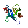
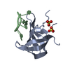
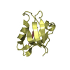
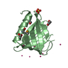
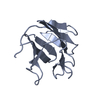
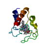
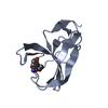
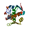
| Title | Complex structure of lysostaphin SH3b domain with peptidoglycan fragment | ||||||
 Components Components |
| ||||||
 Keywords Keywords |  CELL ADHESION / lysostaphin / peptidoglycan / complex / cell wall targeting/binding domain / SH3 CELL ADHESION / lysostaphin / peptidoglycan / complex / cell wall targeting/binding domain / SH3 | ||||||
| Function / homology |  Function and homology informationlysostaphin / cell wall organization / metallopeptidase activity / proteolysis / extracellular region / metal ion binding Function and homology informationlysostaphin / cell wall organization / metallopeptidase activity / proteolysis / extracellular region / metal ion bindingSimilarity search - Function | ||||||
| Biological species |  Staphylococcaceae (Staphylococcus group) Staphylococcaceae (Staphylococcus group) | ||||||
| Method | X-RAY DIFFRACTION / SYNCHROTRON / MOLECULAR REPLACEMENT / Resolution: 1.6 Å | ||||||
 Authors Authors | Jagielska, E. / Nowak, E. / Bochtler, M. / Sabala, I. | ||||||
| Funding support |  Poland, 1items Poland, 1items
| ||||||
 Citation Citation | Journal: To Be Published Title: Complex structure of lysostaphin SH3doamin with peptidoglycan fragment Authors: Jagielska, E. / Nowak, E. / Bochtler, M. / Sabala, I. | ||||||
| History |
|
- Structure visualization
| Structure viewer | Molecule: MolmilJmol/JSmol |
|---|
- Downloads & links
-Download
| PDBx/mmCIF format | 5leo.cif.gz | 100.9 KB | Display | PDBx/mmCIF format |
|---|---|---|---|---|
| PDB format | pdb5leo.ent.gz | 77.3 KB | Display | PDB format |
| PDBx/mmJSON format | 5leo.json.gz | Tree view | PDBx/mmJSON format | |
| Others |  Other downloads Other downloads |
-Validation report
| Arichive directory | https://data.pdbj.org/pub/pdb/validation_reports/le/5leoftp://data.pdbj.org/pub/pdb/validation_reports/le/5leo | HTTPS FTP |
|---|
-Related structure data
| Related structure data |  4lxcS S: Starting model for refinement |
|---|---|
| Similar structure data |


-Links
 PDBj
PDBj- Assembly
| Deposited unit | 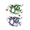
| ||||||||
|---|---|---|---|---|---|---|---|---|---|
| 1 | 
| ||||||||
| 2 | 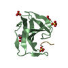
| ||||||||
| Unit cell |
|
-Components
| #1: Protein | / Glycyl-glycine endopeptidase Mass: 10570.948 Da / Num. of mol.: 2 / Fragment: SH3b domain, UNP Residues 401-493 Source method: isolated from a genetically manipulated source Source: (gene. exp.) Staphylococcaceae (Staphylococcus group)Gene: lss / Production host: Escherichia coli (E. coli) / References: UniProt: P10547, lysostaphin#2: Protein/peptide | Mass: 303.274 Da / Num. of mol.: 2 / Source method: obtained synthetically / Source: (synth.) Staphylococcaceae (Staphylococcus group)#3: Chemical | ChemComp-SO4 / Sulfate  Mass: 96.063 Da / Num. of mol.: 6 / Source method: obtained synthetically / Formula: SO4 Mass: 96.063 Da / Num. of mol.: 6 / Source method: obtained synthetically / Formula: SO4#4: Chemical | 2-Methyl-2,4-pentanediol  Mass: 118.174 Da / Num. of mol.: 2 / Source method: obtained synthetically / Formula: C6H14O2 / Comment: precipitant*YM Mass: 118.174 Da / Num. of mol.: 2 / Source method: obtained synthetically / Formula: C6H14O2 / Comment: precipitant*YM#5: Water | ChemComp-HOH / | Water Mass: 18.015 Da / Num. of mol.: 265 / Source method: isolated from a natural source / Formula: H2O Mass: 18.015 Da / Num. of mol.: 265 / Source method: isolated from a natural source / Formula: H2O |
|---|
-Experimental details
-Experiment
| Experiment | Method: X-RAY DIFFRACTION / Number of used crystals: 1 |
|---|
- Sample preparation
| Crystal | Density Matthews: 1.99 Å3/Da / Density % sol: 38.21 % |
|---|---|
| Crystal grow | Temperature: 290 K / Method: vapor diffusion, sitting drop / pH: 8.5 Details: Buffer system 3 (0.1 M Tris (base), Bicine buffer, pH 8.5), 0.09 M NPS (NaNO3, Na2HPO4, (NH4)SO4), 37.5% MPD (racemic), PEG 1000, PEG 3350 |
-Data collection
| Diffraction | Mean temperature: 100 K |
|---|---|
| Diffraction source | Source: SYNCHROTRON / Site: ESRF  / Beamline: ID29 / Wavelength: 0.8726 Å / Beamline: ID29 / Wavelength: 0.8726 Å |
| Detector | Type: DECTRIS PILATUS3 S 6M / Detector: PIXEL / Date: Oct 27, 2014 |
| Radiation | Protocol: SINGLE WAVELENGTH / Monochromatic (M) / Laue (L): M / Scattering type: x-ray |
| Radiation wavelength | Wavelength: 0.8726 Å / Relative weight: 1 |
| Reflection | Resolution: 1.57→60 Å / Num. obs: 25084 / % possible obs: 98.9 % / Redundancy: 4.8 % / Rmerge(I) obs: 0.215 / Net I/σ(I): 5.25 |
- Processing
| Software |
| ||||||||||||||||||||||||||||||||||||||||||||||||||||||||||||||||||||||
|---|---|---|---|---|---|---|---|---|---|---|---|---|---|---|---|---|---|---|---|---|---|---|---|---|---|---|---|---|---|---|---|---|---|---|---|---|---|---|---|---|---|---|---|---|---|---|---|---|---|---|---|---|---|---|---|---|---|---|---|---|---|---|---|---|---|---|---|---|---|---|---|
| Refinement | Method to determine structure: MOLECULAR REPLACEMENT Starting model: 4LXC Resolution: 1.6→39.402 Å / SU ML: 0.2 / Cross valid method: NONE / σ(F): 1.35 / Phase error: 22.79
| ||||||||||||||||||||||||||||||||||||||||||||||||||||||||||||||||||||||
| Solvent computation | Shrinkage radii: 0.9 Å / VDW probe radii: 1.11 Å | ||||||||||||||||||||||||||||||||||||||||||||||||||||||||||||||||||||||
| Refinement step | Cycle: LAST / Resolution: 1.6→39.402 Å
| ||||||||||||||||||||||||||||||||||||||||||||||||||||||||||||||||||||||
| Refine LS restraints |
| ||||||||||||||||||||||||||||||||||||||||||||||||||||||||||||||||||||||
| LS refinement shell |
| ||||||||||||||||||||||||||||||||||||||||||||||||||||||||||||||||||||||
| Refinement TLS params. | Method: refined / Origin x: -27.8405 Å / Origin y: 4.0045 Å / Origin z: 82.5817 Å
| ||||||||||||||||||||||||||||||||||||||||||||||||||||||||||||||||||||||
| Refinement TLS group | Selection details: all |
