 Movie
Movie Controller

[English] 日本語
 Yorodumi
- PDB-5lbz: Structure of the human quinone reductase 2 (NQO2) in complex with... -
+ Open data

- Basic information
| Entry | Database: PDB / ID: 5lbz | ||||||
|---|---|---|---|---|---|---|---|
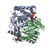
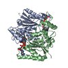
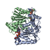
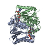
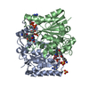
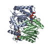
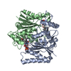
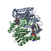
| Title | Structure of the human quinone reductase 2 (NQO2) in complex with pacritinib | ||||||
 Components Components | Ribosyldihydronicotinamide dehydrogenase [quinone] | ||||||
 Keywords Keywords |  OXIDOREDUCTASE / quinone reductase 2 / kinase inhibitor / pancritinib / Ribosyldihydronicotinamide dehydrogenase (NQO2) OXIDOREDUCTASE / quinone reductase 2 / kinase inhibitor / pancritinib / Ribosyldihydronicotinamide dehydrogenase (NQO2) | ||||||
| Function / homology |  Function and homology informationribosyldihydronicotinamide dehydrogenase (quinone) / dihydronicotinamide riboside quinone reductase activity / quinone catabolic process / resveratrol binding / oxidoreductase activity, acting on other nitrogenous compounds as donors / melatonin binding / NAD(P)H dehydrogenase (quinone) activity / Phase I - Functionalization of compounds / chloride ion binding / FAD binding ...ribosyldihydronicotinamide dehydrogenase (quinone) / dihydronicotinamide riboside quinone reductase activity / quinone catabolic process / resveratrol binding / oxidoreductase activity, acting on other nitrogenous compounds as donors / melatonin binding / NAD(P)H dehydrogenase (quinone) activity / Phase I - Functionalization of compounds / chloride ion binding / FAD binding / electron transfer activity / oxidoreductase activity / protein homodimerization activity / extracellular exosome / zinc ion binding / nucleoplasm / cytosol Function and homology informationribosyldihydronicotinamide dehydrogenase (quinone) / dihydronicotinamide riboside quinone reductase activity / quinone catabolic process / resveratrol binding / oxidoreductase activity, acting on other nitrogenous compounds as donors / melatonin binding / NAD(P)H dehydrogenase (quinone) activity / Phase I - Functionalization of compounds / chloride ion binding / FAD binding ...ribosyldihydronicotinamide dehydrogenase (quinone) / dihydronicotinamide riboside quinone reductase activity / quinone catabolic process / resveratrol binding / oxidoreductase activity, acting on other nitrogenous compounds as donors / melatonin binding / NAD(P)H dehydrogenase (quinone) activity / Phase I - Functionalization of compounds / chloride ion binding / FAD binding / electron transfer activity / oxidoreductase activity / protein homodimerization activity / extracellular exosome / zinc ion binding / nucleoplasm / cytosolSimilarity search - Function | ||||||
| Biological species |  Homo sapiens (human) Homo sapiens (human) | ||||||
| Method | X-RAY DIFFRACTION / SYNCHROTRON / FOURIER SYNTHESIS / Resolution: 1.4 Å | ||||||
 Authors Authors | Schneider, S. / Medard, G. / Kuster, B. | ||||||
| Funding support |  Germany, 1items Germany, 1items
| ||||||
 Citation Citation | Journal: Science / Year: 2017 Title: The target landscape of clinical kinase drugs. Authors: Klaeger, S. / Heinzlmeir, S. / Wilhelm, M. / Polzer, H. / Vick, B. / Koenig, P.A. / Reinecke, M. / Ruprecht, B. / Petzoldt, S. / Meng, C. / Zecha, J. / Reiter, K. / Qiao, H. / Helm, D. / ...Authors: Klaeger, S. / Heinzlmeir, S. / Wilhelm, M. / Polzer, H. / Vick, B. / Koenig, P.A. / Reinecke, M. / Ruprecht, B. / Petzoldt, S. / Meng, C. / Zecha, J. / Reiter, K. / Qiao, H. / Helm, D. / Koch, H. / Schoof, M. / Canevari, G. / Casale, E. / Depaolini, S.R. / Feuchtinger, A. / Wu, Z. / Schmidt, T. / Rueckert, L. / Becker, W. / Huenges, J. / Garz, A.K. / Gohlke, B.O. / Zolg, D.P. / Kayser, G. / Vooder, T. / Preissner, R. / Hahne, H. / Tonisson, N. / Kramer, K. / Gotze, K. / Bassermann, F. / Schlegl, J. / Ehrlich, H.C. / Aiche, S. / Walch, A. / Greif, P.A. / Schneider, S. / Felder, E.R. / Ruland, J. / Medard, G. / Jeremias, I. / Spiekermann, K. / Kuster, B. | ||||||
| History |
|
- Structure visualization
| Structure viewer | Molecule: MolmilJmol/JSmol |
|---|
- Downloads & links
-Download
| PDBx/mmCIF format | 5lbz.cif.gz | 225.6 KB | Display | PDBx/mmCIF format |
|---|---|---|---|---|
| PDB format | pdb5lbz.ent.gz | 177.9 KB | Display | PDB format |
| PDBx/mmJSON format | 5lbz.json.gz | Tree view | PDBx/mmJSON format | |
| Others |  Other downloads Other downloads |
-Validation report
| Arichive directory | https://data.pdbj.org/pub/pdb/validation_reports/lb/5lbzftp://data.pdbj.org/pub/pdb/validation_reports/lb/5lbz | HTTPS FTP |
|---|
-Related structure data
| Related structure data | 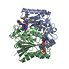 5lbwC  5lbyC  5m5aC  5mafC 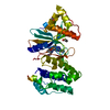 5magC  5mahC 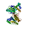 5maiC 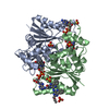 5lbuS S: Starting model for refinement C: citing same article ( |
|---|---|
| Similar structure data |


-Links
 PDBj
PDBj- Assembly
| Deposited unit | 
| ||||||||||||||||||
|---|---|---|---|---|---|---|---|---|---|---|---|---|---|---|---|---|---|---|---|
| 1 |
| ||||||||||||||||||
| Unit cell |
| ||||||||||||||||||
| Noncrystallographic symmetry (NCS) | NCS domain:
NCS domain segments: Component-ID: 0 / Ens-ID: 1 / Beg auth comp-ID: LYS / Beg label comp-ID: LYS / End auth comp-ID: GLY / End label comp-ID: GLY / Refine code: 0 / Auth seq-ID: 3 - 229 / Label seq-ID: 4 - 230
|
-Components
-Protein , 1 types, 2 molecules AB
| #1: Protein | Mass: 26775.398 Da / Num. of mol.: 2 Source method: isolated from a genetically manipulated source Source: (gene. exp.) Homo sapiens (human) / Gene: NQO2, NMOR2 / Plasmid: pET28a / Production host:  Escherichia coli (E. coli) Escherichia coli (E. coli)References: UniProt: P16083, ribosyldihydronicotinamide dehydrogenase (quinone) |
|---|
-Non-polymers , 5 types, 437 molecules 


| #2: Chemical | Flavin adenine dinucleotide Mass: 785.550 Da / Num. of mol.: 2 / Source method: obtained synthetically / Formula: C27H33N9O15P2 / Comment: FAD*YM Mass: 785.550 Da / Num. of mol.: 2 / Source method: obtained synthetically / Formula: C27H33N9O15P2 / Comment: FAD*YM#3: Chemical | Pacritinib Mass: 472.579 Da / Num. of mol.: 2 / Source method: obtained synthetically / Formula: C28H32N4O3 / Comment: medication, anticancer, inhibitor*YM Mass: 472.579 Da / Num. of mol.: 2 / Source method: obtained synthetically / Formula: C28H32N4O3 / Comment: medication, anticancer, inhibitor*YM#4: Chemical | ChemComp-SO4 / Sulfate Mass: 96.063 Da / Num. of mol.: 4 / Source method: obtained synthetically / Formula: SO4 Mass: 96.063 Da / Num. of mol.: 4 / Source method: obtained synthetically / Formula: SO4#5: Chemical |  Mass: 65.409 Da / Num. of mol.: 2 / Source method: obtained synthetically / Formula: Zn Mass: 65.409 Da / Num. of mol.: 2 / Source method: obtained synthetically / Formula: Zn#6: Water | ChemComp-HOH / | WaterMass: 18.015 Da / Num. of mol.: 427 / Source method: isolated from a natural source / Formula: H2O |
|---|
-Experimental details
-Experiment
| Experiment | Method: X-RAY DIFFRACTION / Number of used crystals: 1 |
|---|
- Sample preparation
| Crystal | Density Matthews: 2.41 Å3/Da / Density % sol: 49.06 % |
|---|---|
| Crystal grow | Temperature: 293 K / Method: vapor diffusion / Details: 200 mM sodium sulphate, 2.2 M ammonium sulphate |
-Data collection
| Diffraction | Mean temperature: 100 K |
|---|---|
| Diffraction source | Source: SYNCHROTRON / Site: SLS  / Beamline: X06SA / Wavelength: 0.9785 Å / Beamline: X06SA / Wavelength: 0.9785 Å |
| Detector | Type: DECTRIS EIGER X 16M / Detector: PIXEL / Date: Nov 30, 2015 |
| Radiation | Protocol: SINGLE WAVELENGTH / Monochromatic (M) / Laue (L): M / Scattering type: x-ray |
| Radiation wavelength | Wavelength: 0.9785 Å / Relative weight: 1 |
| Reflection | Resolution: 1.4→48.6 Å / Num. obs: 97699 / % possible obs: 95 % / Redundancy: 4.5 % / CC1/2: 0.999 / Rmerge(I) obs: 0.045 / Net I/σ(I): 21.11 |
| Reflection shell | Resolution: 1.4→1.45 Å / Redundancy: 4.4 % / Rmerge(I) obs: 0.329 / Mean I/σ(I) obs: 5.3 / % possible all: 91 |
- Processing
| Software |
| ||||||||||||||||||||||||||||||||||||||||||||||||||||||||||||||||||||||||||||||||||||||||||||||||||||||||||||||||||||||||||||||||||||||||||||||||||||||||||||||||||||||||||||||||||||||
|---|---|---|---|---|---|---|---|---|---|---|---|---|---|---|---|---|---|---|---|---|---|---|---|---|---|---|---|---|---|---|---|---|---|---|---|---|---|---|---|---|---|---|---|---|---|---|---|---|---|---|---|---|---|---|---|---|---|---|---|---|---|---|---|---|---|---|---|---|---|---|---|---|---|---|---|---|---|---|---|---|---|---|---|---|---|---|---|---|---|---|---|---|---|---|---|---|---|---|---|---|---|---|---|---|---|---|---|---|---|---|---|---|---|---|---|---|---|---|---|---|---|---|---|---|---|---|---|---|---|---|---|---|---|---|---|---|---|---|---|---|---|---|---|---|---|---|---|---|---|---|---|---|---|---|---|---|---|---|---|---|---|---|---|---|---|---|---|---|---|---|---|---|---|---|---|---|---|---|---|---|---|---|---|
| Refinement | Method to determine structure: FOURIER SYNTHESIS Starting model: 5LBU Resolution: 1.4→48.6 Å / Cor.coef. Fo:Fc: 0.98 / Cor.coef. Fo:Fc free: 0.975 / SU B: 1.146 / SU ML: 0.021 / Cross valid method: THROUGHOUT / ESU R: 0.044 / ESU R Free: 0.041 / Details: HYDROGENS HAVE BEEN ADDED IN THE RIDING POSITIONS
| ||||||||||||||||||||||||||||||||||||||||||||||||||||||||||||||||||||||||||||||||||||||||||||||||||||||||||||||||||||||||||||||||||||||||||||||||||||||||||||||||||||||||||||||||||||||
| Solvent computation | Ion probe radii: 0.8 Å / Shrinkage radii: 0.8 Å / VDW probe radii: 1.4 Å | ||||||||||||||||||||||||||||||||||||||||||||||||||||||||||||||||||||||||||||||||||||||||||||||||||||||||||||||||||||||||||||||||||||||||||||||||||||||||||||||||||||||||||||||||||||||
| Displacement parameters | Biso mean: 14.062 Å2
| ||||||||||||||||||||||||||||||||||||||||||||||||||||||||||||||||||||||||||||||||||||||||||||||||||||||||||||||||||||||||||||||||||||||||||||||||||||||||||||||||||||||||||||||||||||||
| Refinement step | Cycle: LAST / Resolution: 1.4→48.6 Å
| ||||||||||||||||||||||||||||||||||||||||||||||||||||||||||||||||||||||||||||||||||||||||||||||||||||||||||||||||||||||||||||||||||||||||||||||||||||||||||||||||||||||||||||||||||||||
| Refine LS restraints |
|