 Movie
Movie Controller


+ Open data

- Basic information
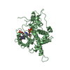
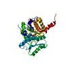
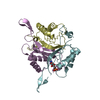
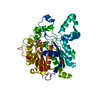
| Entry | Database: PDB / ID: 5l2x | ||||||
|---|---|---|---|---|---|---|---|
| Title | Crystal structure of human PrimPol ternary complex | ||||||
 Components Components |
| ||||||
 Keywords Keywords | TRANSFERASE/DNA /  Protein-DNA complex / TRANSFERASE-DNA complex Protein-DNA complex / TRANSFERASE-DNA complex | ||||||
| Function / homology |  Function and homology information Function and homology informationDNA primase AEP / R-loop processing / mitochondrial DNA replication / DNA primase activity / mitochondrial DNA repair / replication fork processing / translesion synthesis / error-prone translesion synthesis / response to UV / DNA-directed RNA polymerase complex ...DNA primase AEP / R-loop processing / mitochondrial DNA replication / DNA primase activity / mitochondrial DNA repair / replication fork processing / translesion synthesis / error-prone translesion synthesis / response to UV / DNA-directed RNA polymerase complex / replication fork / manganese ion binding / DNA-directed DNA polymerase / DNA-directed DNA polymerase activity / mitochondrial matrix / chromatin binding / zinc ion binding / nucleusSimilarity search - Function | ||||||
| Biological species |  Homo sapiens (human) Homo sapiens (human) | ||||||
| Method | X-RAY DIFFRACTION / SYNCHROTRON / SAD / Resolution: 2.2 Å | ||||||
 Authors Authors | Rechkoblit, O. / Gupta, Y.K. / Malik, R. / Rajashankar, K.R. / Johnson, R.E. / Prakash, L. / Prakash, S. / Aggarwal, A.K. | ||||||
 Citation Citation | Journal: Sci Adv / Year: 2016 Title: Structure and mechanism of human PrimPol, a DNA polymerase with primase activity. Authors: Rechkoblit, O. / Gupta, Y.K. / Malik, R. / Rajashankar, K.R. / Johnson, R.E. / Prakash, L. / Prakash, S. / Aggarwal, A.K. | ||||||
| History |
|
- Structure visualization
| Structure viewer | Molecule: MolmilJmol/JSmol |
|---|
- Downloads & links
-Download
| PDBx/mmCIF format | 5l2x.cif.gz | 146.8 KB | Display | PDBx/mmCIF format |
|---|---|---|---|---|
| PDB format | pdb5l2x.ent.gz | 109.6 KB | Display | PDB format |
| PDBx/mmJSON format | 5l2x.json.gz | Tree view | PDBx/mmJSON format | |
| Others |  Other downloads Other downloads |
-Validation report
| Arichive directory | https://data.pdbj.org/pub/pdb/validation_reports/l2/5l2xftp://data.pdbj.org/pub/pdb/validation_reports/l2/5l2x | HTTPS FTP |
|---|
-Related structure data
| Similar structure data |
|---|


-Links
 PDBj
PDBj


- Assembly
| Deposited unit | 
| ||||||||
|---|---|---|---|---|---|---|---|---|---|
| 1 | 
| ||||||||
| 2 | 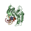
| ||||||||
| Unit cell |
|
-Components
-Protein , 1 types, 2 molecules AB
| #1: Protein | Mass: 40909.309 Da / Num. of mol.: 2 Source method: isolated from a genetically manipulated source Source: (gene. exp.) Homo sapiens (human) / Gene: PRIMPOL, CCDC111 / Production host:  Saccharomyces cerevisiae (brewer's yeast) Saccharomyces cerevisiae (brewer's yeast)References: UniProt: Q96LW4, Transferases; Transferring phosphorus-containing groups; Nucleotidyltransferases |
|---|
-DNA chain , 2 types, 4 molecules CGDH
| #2: DNA chain | Mass: 5149.148 Da / Num. of mol.: 2 / Source method: obtained synthetically / Source: (synth.) Homo sapiens (human)#3: DNA chain | Mass: 4087.657 Da / Num. of mol.: 2 / Source method: obtained synthetically / Source: (synth.) Homo sapiens (human) |
|---|
-Non-polymers , 3 types, 102 molecules 


| #4: Chemical | Deoxyadenosine triphosphate Mass: 491.182 Da / Num. of mol.: 2 / Source method: obtained synthetically / Formula: C10H16N5O12P3 Mass: 491.182 Da / Num. of mol.: 2 / Source method: obtained synthetically / Formula: C10H16N5O12P3#5: Chemical |  Mass: 40.078 Da / Num. of mol.: 2 / Source method: obtained synthetically / Formula: Ca Mass: 40.078 Da / Num. of mol.: 2 / Source method: obtained synthetically / Formula: Ca#6: Water | ChemComp-HOH / | WaterMass: 18.015 Da / Num. of mol.: 98 / Source method: isolated from a natural source / Formula: H2O |
|---|
-Experimental details
-Experiment
| Experiment | Method: X-RAY DIFFRACTION / Number of used crystals: 1 |
|---|
- Sample preparation
| Crystal | Density Matthews: 2.22 Å3/Da / Density % sol: 50.47 % |
|---|---|
| Crystal grow | Temperature: 293 K / Method: vapor diffusion, sitting drop / pH: 7.5 / Details: 17-23% PEG 3350 |
-Data collection
| Diffraction | Mean temperature: 100 K |
|---|---|
| Diffraction source | Source: SYNCHROTRON / Site: APS  / Beamline: 24-ID-C / Wavelength: 0.9792 Å / Beamline: 24-ID-C / Wavelength: 0.9792 Å |
| Detector | Type: DECTRIS PILATUS3 S 6M / Detector: PIXEL / Date: Feb 8, 2016 / Details: MD2 |
| Radiation | Monochromator: Si(111) / Protocol: SINGLE WAVELENGTH / Monochromatic (M) / Laue (L): M / Scattering type: x-ray |
| Radiation wavelength | Wavelength: 0.9792 Å / Relative weight: 1 |
| Reflection | Resolution: 2.2→39.354 Å / Num. obs: 38848 / % possible obs: 94.2 % / Redundancy: 2.5 % / Rsym value: 0.01 / Net I/σ(I): 6.2 |
- Processing
| Software |
| ||||||||||||||||||||||||||||||||||||||||||||||||||||||||||||||||||||||||||||||||||||||||||||||||||
|---|---|---|---|---|---|---|---|---|---|---|---|---|---|---|---|---|---|---|---|---|---|---|---|---|---|---|---|---|---|---|---|---|---|---|---|---|---|---|---|---|---|---|---|---|---|---|---|---|---|---|---|---|---|---|---|---|---|---|---|---|---|---|---|---|---|---|---|---|---|---|---|---|---|---|---|---|---|---|---|---|---|---|---|---|---|---|---|---|---|---|---|---|---|---|---|---|---|---|---|
| Refinement | Method to determine structure: SAD / Resolution: 2.2→39.354 Å / SU ML: 0.32 / Cross valid method: FREE R-VALUE / σ(F): 1.98 / Phase error: 29.63 / Stereochemistry target values: ML
| ||||||||||||||||||||||||||||||||||||||||||||||||||||||||||||||||||||||||||||||||||||||||||||||||||
| Solvent computation | Shrinkage radii: 0.9 Å / VDW probe radii: 1.11 Å / Solvent model: FLAT BULK SOLVENT MODEL | ||||||||||||||||||||||||||||||||||||||||||||||||||||||||||||||||||||||||||||||||||||||||||||||||||
| Refinement step | Cycle: LAST / Resolution: 2.2→39.354 Å
| ||||||||||||||||||||||||||||||||||||||||||||||||||||||||||||||||||||||||||||||||||||||||||||||||||
| Refine LS restraints |
| ||||||||||||||||||||||||||||||||||||||||||||||||||||||||||||||||||||||||||||||||||||||||||||||||||
| LS refinement shell |
|
