 Movie
Movie Controller

[English] 日本語
 Yorodumi
- PDB-5l21: Crystal structure of BoNT/A receptor binding domain in complex wi... -
+ Open data

- Basic information
| Entry | Database: PDB / ID: 5l21 | ||||||
|---|---|---|---|---|---|---|---|
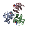
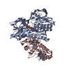
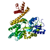
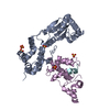
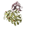
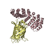
| Title | Crystal structure of BoNT/A receptor binding domain in complex with VHH C2 | ||||||
 Components Components |
| ||||||
 Keywords Keywords |  TOXIN / Neutralizing antibody / VHH / Botulinum neurotoxin / BoNT-A-VHH complex TOXIN / Neutralizing antibody / VHH / Botulinum neurotoxin / BoNT-A-VHH complex | ||||||
| Function / homology |  Function and homology information Function and homology informationhost cell junction / negative regulation of neurotransmitter secretion / bontoxilysin / host cell presynaptic membrane / host cell cytoplasmic vesicle / host cell cytosol / protein transmembrane transporter activity / metalloendopeptidase activity / toxin activity / membrane => GO:0016020 ...host cell junction / negative regulation of neurotransmitter secretion / bontoxilysin / host cell presynaptic membrane / host cell cytoplasmic vesicle / host cell cytosol / protein transmembrane transporter activity / metalloendopeptidase activity / toxin activity / membrane => GO:0016020 / host cell plasma membrane / proteolysis / zinc ion binding / extracellular region / membraneSimilarity search - Function | ||||||
| Biological species |   Clostridium botulinum (bacteria) Clostridium botulinum (bacteria) Vicugna pacos (alpaca) Vicugna pacos (alpaca) | ||||||
| Method | X-RAY DIFFRACTION / SYNCHROTRON / MOLECULAR REPLACEMENT / Resolution: 1.68 Å | ||||||
 Authors Authors | Yao, G. / Jin, R. | ||||||
| Funding support |  United States, 1items United States, 1items
| ||||||
 Citation Citation | Journal: Sci Rep / Year: 2017 Title: A camelid single-domain antibody neutralizes botulinum neurotoxin A by blocking host receptor binding. Authors: Yao, G. / Lam, K.H. / Weisemann, J. / Peng, L. / Krez, N. / Perry, K. / Shoemaker, C.B. / Dong, M. / Rummel, A. / Jin, R. | ||||||
| History |
|
- Structure visualization
| Structure viewer | Molecule: MolmilJmol/JSmol |
|---|
- Downloads & links
-Download
| PDBx/mmCIF format | 5l21.cif.gz | 249.5 KB | Display | PDBx/mmCIF format |
|---|---|---|---|---|
| PDB format | pdb5l21.ent.gz | 197.5 KB | Display | PDB format |
| PDBx/mmJSON format | 5l21.json.gz | Tree view | PDBx/mmJSON format | |
| Others |  Other downloads Other downloads |
-Validation report
| Arichive directory | https://data.pdbj.org/pub/pdb/validation_reports/l2/5l21ftp://data.pdbj.org/pub/pdb/validation_reports/l2/5l21 | HTTPS FTP |
|---|
-Related structure data
| Related structure data | 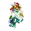 3fuoS S: Starting model for refinement |
|---|---|
| Similar structure data |


-Links
 PDBj
PDBj


- Assembly
| Deposited unit | 
| ||||||||
|---|---|---|---|---|---|---|---|---|---|
| 1 |
| ||||||||
| Unit cell |
|
-Components
| #1: Protein | Mass: 49877.488 Da / Num. of mol.: 1 / Mutation: T1158A Source method: isolated from a genetically manipulated source Source: (gene. exp.) Clostridium botulinum (strain Hall / ATCC 3502 / NCTC 13319 / Type A) (bacteria)Strain: Hall / ATCC 3502 / NCTC 13319 / Type A / Gene: botA, CBO0806, CLC_0862 / Production host: Escherichia coli (E. coli)References: UniProt: A5HZZ9, UniProt: P0DPI1*PLUS, bontoxilysin |
|---|---|
| #2: Antibody | Mass: 13326.828 Da / Num. of mol.: 1 Source method: isolated from a genetically manipulated source Source: (gene. exp.) Vicugna pacos (alpaca) / Production host: Escherichia coli (E. coli) |
| #3: Water | ChemComp-HOH / Water Mass: 18.015 Da / Num. of mol.: 566 / Source method: isolated from a natural source / Formula: H2O Mass: 18.015 Da / Num. of mol.: 566 / Source method: isolated from a natural source / Formula: H2O |
-Experimental details
-Experiment
| Experiment | Method: X-RAY DIFFRACTION / Number of used crystals: 1 |
|---|
- Sample preparation
| Crystal | Density Matthews: 2.65 Å3/Da / Density % sol: 53.65 % |
|---|---|
| Crystal grow | Temperature: 291 K / Method: vapor diffusion, hanging drop Details: 18% polyethylene glycol 6,000; 100 mM Sodium Acetate, pH 5.0 |
-Data collection
| Diffraction | Mean temperature: 100 K |
|---|---|
| Diffraction source | Source: SYNCHROTRON / Site: APS / Beamline: 24-ID-C / Wavelength: 0.97918 Å |
| Detector | Type: ADSC QUANTUM 315r / Detector: CCD / Date: Mar 12, 2012 |
| Radiation | Protocol: SINGLE WAVELENGTH / Monochromatic (M) / Laue (L): M / Scattering type: x-ray |
| Radiation wavelength | Wavelength: 0.97918 Å / Relative weight: 1 |
| Reflection | Resolution: 1.68→35.99 Å / Num. obs: 74608 / % possible obs: 99.5 % / Redundancy: 2.9 % / CC1/2: 0.998 / Rmerge(I) obs: 0.041 / Net I/σ(I): 12.8 |
| Reflection shell | Resolution: 1.68→1.71 Å / Redundancy: 2.9 % / Rmerge(I) obs: 0.414 / Mean I/σ(I) obs: 2.3 / CC1/2: 0.843 / % possible all: 99.7 |
- Processing
| Software |
| ||||||||||||||||||||||||||||||||||||||||||||||||||||||||||||||||||||||||||||||||||||||||||||||||||||||||||||||||||||||||||||||||||||||||||||||||||||||||||||||||||||||||||||||||||||||||||||||||||||||||||||||||||||||||||||||||||||||||||||||||||||||||||
|---|---|---|---|---|---|---|---|---|---|---|---|---|---|---|---|---|---|---|---|---|---|---|---|---|---|---|---|---|---|---|---|---|---|---|---|---|---|---|---|---|---|---|---|---|---|---|---|---|---|---|---|---|---|---|---|---|---|---|---|---|---|---|---|---|---|---|---|---|---|---|---|---|---|---|---|---|---|---|---|---|---|---|---|---|---|---|---|---|---|---|---|---|---|---|---|---|---|---|---|---|---|---|---|---|---|---|---|---|---|---|---|---|---|---|---|---|---|---|---|---|---|---|---|---|---|---|---|---|---|---|---|---|---|---|---|---|---|---|---|---|---|---|---|---|---|---|---|---|---|---|---|---|---|---|---|---|---|---|---|---|---|---|---|---|---|---|---|---|---|---|---|---|---|---|---|---|---|---|---|---|---|---|---|---|---|---|---|---|---|---|---|---|---|---|---|---|---|---|---|---|---|---|---|---|---|---|---|---|---|---|---|---|---|---|---|---|---|---|---|---|---|---|---|---|---|---|---|---|---|---|---|---|---|---|---|---|---|---|---|---|---|---|---|---|---|---|---|---|---|---|---|
| Refinement | Method to determine structure: MOLECULAR REPLACEMENT Starting model: 3FUO Resolution: 1.68→35.99 Å / SU ML: 0.17 / Cross valid method: FREE R-VALUE / σ(F): 1.34 / Phase error: 21.11
| ||||||||||||||||||||||||||||||||||||||||||||||||||||||||||||||||||||||||||||||||||||||||||||||||||||||||||||||||||||||||||||||||||||||||||||||||||||||||||||||||||||||||||||||||||||||||||||||||||||||||||||||||||||||||||||||||||||||||||||||||||||||||||
| Solvent computation | Shrinkage radii: 0.9 Å / VDW probe radii: 1.11 Å | ||||||||||||||||||||||||||||||||||||||||||||||||||||||||||||||||||||||||||||||||||||||||||||||||||||||||||||||||||||||||||||||||||||||||||||||||||||||||||||||||||||||||||||||||||||||||||||||||||||||||||||||||||||||||||||||||||||||||||||||||||||||||||
| Refinement step | Cycle: LAST / Resolution: 1.68→35.99 Å
| ||||||||||||||||||||||||||||||||||||||||||||||||||||||||||||||||||||||||||||||||||||||||||||||||||||||||||||||||||||||||||||||||||||||||||||||||||||||||||||||||||||||||||||||||||||||||||||||||||||||||||||||||||||||||||||||||||||||||||||||||||||||||||
| Refine LS restraints |
| ||||||||||||||||||||||||||||||||||||||||||||||||||||||||||||||||||||||||||||||||||||||||||||||||||||||||||||||||||||||||||||||||||||||||||||||||||||||||||||||||||||||||||||||||||||||||||||||||||||||||||||||||||||||||||||||||||||||||||||||||||||||||||
| LS refinement shell |
| ||||||||||||||||||||||||||||||||||||||||||||||||||||||||||||||||||||||||||||||||||||||||||||||||||||||||||||||||||||||||||||||||||||||||||||||||||||||||||||||||||||||||||||||||||||||||||||||||||||||||||||||||||||||||||||||||||||||||||||||||||||||||||
| Refinement TLS params. | Method: refined / Refine-ID: X-RAY DIFFRACTION
| ||||||||||||||||||||||||||||||||||||||||||||||||||||||||||||||||||||||||||||||||||||||||||||||||||||||||||||||||||||||||||||||||||||||||||||||||||||||||||||||||||||||||||||||||||||||||||||||||||||||||||||||||||||||||||||||||||||||||||||||||||||||||||
| Refinement TLS group |
|
