 Movie
Movie Controller

[English] 日本語
 Yorodumi
- PDB-5l0a: Human muscle fructose-1,6-bisphosphatase E69Q mutant in active R-... -
+ Open data

- Basic information
| Entry | Database: PDB / ID: 5l0a | ||||||
|---|---|---|---|---|---|---|---|
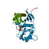
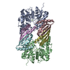
| Title | Human muscle fructose-1,6-bisphosphatase E69Q mutant in active R-state in complex with fructose-1,6-bisphosphate | ||||||
 Components Components | Fructose-1,6-bisphosphatase isozyme 2 | ||||||
 Keywords Keywords |  HYDROLASE / carbohydrate metabolism / glyconeogenesis / muscle izoenzyme / FBPase / R-state / Leucine lock / E69Q / fructose-1 / 6-bisphosphate HYDROLASE / carbohydrate metabolism / glyconeogenesis / muscle izoenzyme / FBPase / R-state / Leucine lock / E69Q / fructose-1 / 6-bisphosphate | ||||||
| Function / homology |  Function and homology information Function and homology informationsucrose biosynthetic process / fructose-bisphosphatase / fructose 1,6-bisphosphate 1-phosphatase activity / fructose 1,6-bisphosphate metabolic process / fructose 6-phosphate metabolic process / Gluconeogenesis / fructose metabolic process / anchoring junction / gluconeogenesis / Z disc ...sucrose biosynthetic process / fructose-bisphosphatase / fructose 1,6-bisphosphate 1-phosphatase activity / fructose 1,6-bisphosphate metabolic process / fructose 6-phosphate metabolic process / Gluconeogenesis / fructose metabolic process / anchoring junction / gluconeogenesis / Z disc / extracellular exosome / identical protein binding / metal ion binding / nucleus / plasma membrane / cytosol / cytoplasmSimilarity search - Function | ||||||
| Biological species |  Homo sapiens (human) Homo sapiens (human) | ||||||
| Method | X-RAY DIFFRACTION / SYNCHROTRON / MOLECULAR REPLACEMENT / Resolution: 2.302 Å | ||||||
 Authors Authors | Barciszewski, J. / Wisniewski, J. / Kolodziejczyk, R. / Dzugaj, A. / Jaskolski, M. / Rakus, D. | ||||||
| Funding support |  Poland, 1items Poland, 1items
| ||||||
 Citation Citation | Journal: To Be Published Title: Structural studies of human muscle FBPase Authors: Barciszewski, J. / Szpotkowski, K. / Wisniewski, J. / Kolodziejczyk, R. / Jaskolski, M. / Rakus, D. / Dzugaj, A. #1: Journal: Acta Crystallogr. D Biol. Crystallogr. / Year: 2011Title: Structure of E69Q mutant of human muscle fructose-1,6-bisphosphatase. Authors: Zarzycki, M. / Kolodziejczyk, R. / Maciaszczyk-Dziubinska, E. / Wysocki, R. / Jaskolski, M. / Dzugaj, A. #2: Journal: PLoS ONE / Year: 2013Title: Crystal structures of human muscle fructose-1,6-bisphosphatase: novel quaternary states, enhanced AMP affinity, and allosteric signal transmission pathway. Authors: Shi, R. / Chen, Z.Y. / Zhu, D.W. / Li, C. / Shan, Y. / Xu, G. / Lin, S.X. #3: Journal: Acta Crystallogr D Struct Biol / Year: 2016Title: T-to-R switch of muscle fructose-1,6-bisphosphatase involves fundamental changes of secondary and quaternary structure. Authors: Barciszewski, J. / Wisniewski, J. / Kolodziejczyk, R. / Jaskolski, M. / Rakus, D. / Dzugaj, A. | ||||||
| History |
|
- Structure visualization
| Structure viewer | Molecule: MolmilJmol/JSmol |
|---|
- Downloads & links
-Download
| PDBx/mmCIF format | 5l0a.cif.gz | 170.5 KB | Display | PDBx/mmCIF format |
|---|---|---|---|---|
| PDB format | pdb5l0a.ent.gz | 138.7 KB | Display | PDB format |
| PDBx/mmJSON format | 5l0a.json.gz | Tree view | PDBx/mmJSON format | |
| Others |  Other downloads Other downloads |
-Validation report
| Arichive directory | https://data.pdbj.org/pub/pdb/validation_reports/l0/5l0aftp://data.pdbj.org/pub/pdb/validation_reports/l0/5l0a | HTTPS FTP |
|---|
-Related structure data
| Related structure data |  5k54C  5k55C  5k56C  5et5S S: Starting model for refinement C: citing same article ( |
|---|---|
| Similar structure data | |
| Experimental dataset #1 | Data reference: 10.18150/repod.3968588 / Data set type: diffraction image data |


-Links
 PDBj
PDBj

- Assembly
| Deposited unit | 
| ||||||||
|---|---|---|---|---|---|---|---|---|---|
| 1 | 
| ||||||||
| Unit cell |
|
-Components
| #1: Protein | Mass: 36665.910 Da / Num. of mol.: 1 Source method: isolated from a genetically manipulated source Details: Gaps in the sequence indicate residues that were not modeled because of poor electron density Source: (gene. exp.) Homo sapiens (human) / Tissue: skeletal muscle / Gene: FBP2 / Plasmid: pkk223-3 / Production host:  Escherichia coli (E. coli) / Strain (production host): C100 / References: UniProt: O00757, fructose-bisphosphatase Escherichia coli (E. coli) / Strain (production host): C100 / References: UniProt: O00757, fructose-bisphosphatase |
|---|---|
| #2: Sugar | ChemComp-FBP / Fructose 1,6-bisphosphate  Type: D-saccharide, beta linking / Mass: 340.116 Da / Num. of mol.: 1 Type: D-saccharide, beta linking / Mass: 340.116 Da / Num. of mol.: 1Source method: isolated from a genetically manipulated source Formula: C6H14O12P2 |
| #3: Water | ChemComp-HOH / Water Mass: 18.015 Da / Num. of mol.: 12 / Source method: isolated from a natural source / Formula: H2O Mass: 18.015 Da / Num. of mol.: 12 / Source method: isolated from a natural source / Formula: H2O |
-Experimental details
-Experiment
| Experiment | Method: X-RAY DIFFRACTION / Number of used crystals: 1 |
|---|
- Sample preparation
| Crystal | Density Matthews: 2.09 Å3/Da / Density % sol: 41.23 % |
|---|---|
| Crystal grow | Temperature: 292 K / Method: vapor diffusion, hanging drop / pH: 7.4 Details: 10mM magnesium chloride, 2M sodium chloride, 10%PEG6000, 10mM Tris |
-Data collection
| Diffraction | Mean temperature: 100 K |
|---|---|
| Diffraction source | Source: SYNCHROTRON / Site: BESSY  / Beamline: 14.3 / Wavelength: 0.8943 Å / Beamline: 14.3 / Wavelength: 0.8943 Å |
| Detector | Type: RAYONIX SX-165mm / Detector: CCD / Date: Jun 27, 2010 |
| Radiation | Monochromator: Double Crystal Monochromator, Si-111 crystal / Protocol: SINGLE WAVELENGTH / Monochromatic (M) / Laue (L): M / Scattering type: x-ray |
| Radiation wavelength | Wavelength: 0.8943 Å / Relative weight: 1 |
| Reflection | Resolution: 2.3→31.03 Å / Num. obs: 13949 / % possible obs: 93.9 % / Redundancy: 8.7 % / Rmerge(I) obs: 0.076 / Net I/σ(I): 3.49 |
| Reflection shell | Resolution: 2.3→2.38 Å / Redundancy: 5.7 % / Rmerge(I) obs: 0.622 / Mean I/σ(I) obs: 2 / % possible all: 69.5 |
- Processing
| Software |
| ||||||||||||||||||||||||||||||||||||||||||||||||||||||||||||||||||||||||||||||||||||||||||||||||||||
|---|---|---|---|---|---|---|---|---|---|---|---|---|---|---|---|---|---|---|---|---|---|---|---|---|---|---|---|---|---|---|---|---|---|---|---|---|---|---|---|---|---|---|---|---|---|---|---|---|---|---|---|---|---|---|---|---|---|---|---|---|---|---|---|---|---|---|---|---|---|---|---|---|---|---|---|---|---|---|---|---|---|---|---|---|---|---|---|---|---|---|---|---|---|---|---|---|---|---|---|---|---|
| Refinement | Method to determine structure: MOLECULAR REPLACEMENT Starting model: 5ET5 Resolution: 2.302→31.027 Å / SU ML: 0.31 / Cross valid method: FREE R-VALUE / σ(F): 1.33 / Phase error: 32.44 / Stereochemistry target values: ML
| ||||||||||||||||||||||||||||||||||||||||||||||||||||||||||||||||||||||||||||||||||||||||||||||||||||
| Solvent computation | Shrinkage radii: 0.9 Å / VDW probe radii: 1.11 Å / Solvent model: FLAT BULK SOLVENT MODEL | ||||||||||||||||||||||||||||||||||||||||||||||||||||||||||||||||||||||||||||||||||||||||||||||||||||
| Refinement step | Cycle: LAST / Resolution: 2.302→31.027 Å
| ||||||||||||||||||||||||||||||||||||||||||||||||||||||||||||||||||||||||||||||||||||||||||||||||||||
| Refine LS restraints |
| ||||||||||||||||||||||||||||||||||||||||||||||||||||||||||||||||||||||||||||||||||||||||||||||||||||
| LS refinement shell |
| ||||||||||||||||||||||||||||||||||||||||||||||||||||||||||||||||||||||||||||||||||||||||||||||||||||
| Refinement TLS params. | Method: refined / Refine-ID: X-RAY DIFFRACTION
| ||||||||||||||||||||||||||||||||||||||||||||||||||||||||||||||||||||||||||||||||||||||||||||||||||||
| Refinement TLS group |
|
