 Movie
Movie Controller

[English] 日本語
 Yorodumi
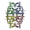
- PDB-5et6: Human muscle fructose-1,6-bisphosphatase in inactive T-state in c... -
+ Open data

- Basic information
| Entry | Database: PDB / ID: 5et6 | ||||||
|---|---|---|---|---|---|---|---|
| Title | Human muscle fructose-1,6-bisphosphatase in inactive T-state in complex with AMP | ||||||
 Components Components | Fructose-1,6-bisphosphatase isozyme 2 | ||||||
 Keywords Keywords |  HYDROLASE / carbohydrate metabolism / glyconeogenesis / muscle / FBPase / T-state / AMP HYDROLASE / carbohydrate metabolism / glyconeogenesis / muscle / FBPase / T-state / AMP | ||||||
| Function / homology |  Function and homology information Function and homology informationsucrose biosynthetic process / fructose-bisphosphatase / fructose 1,6-bisphosphate 1-phosphatase activity / fructose 6-phosphate metabolic process / Gluconeogenesis / fructose metabolic process / fructose 1,6-bisphosphate metabolic process / anchoring junction / gluconeogenesis / Z disc ...sucrose biosynthetic process / fructose-bisphosphatase / fructose 1,6-bisphosphate 1-phosphatase activity / fructose 6-phosphate metabolic process / Gluconeogenesis / fructose metabolic process / fructose 1,6-bisphosphate metabolic process / anchoring junction / gluconeogenesis / Z disc / extracellular exosome / identical protein binding / metal ion binding / nucleus / plasma membrane / cytosol / cytoplasmSimilarity search - Function | ||||||
| Biological species |  Homo sapiens (human) Homo sapiens (human) | ||||||
| Method | X-RAY DIFFRACTION / SYNCHROTRON / MOLECULAR REPLACEMENT / Resolution: 1.845 Å | ||||||
 Authors Authors | Barciszewski, J. / Wisniewski, J. / Kolodziejczyk, R. / Dzugaj, A. / Jaskolski, M. / Rakus, D. | ||||||
| Funding support |  Poland, 1items Poland, 1items
| ||||||
 Citation Citation | Journal: Acta Crystallogr D Struct Biol / Year: 2016 Title: T-to-R switch of muscle fructose-1,6-bisphosphatase involves fundamental changes of secondary and quaternary structure. Authors: Barciszewski, J. / Wisniewski, J. / Kolodziejczyk, R. / Jaskolski, M. / Rakus, D. / Dzugaj, A. #1: Journal: Acta Crystallogr. D Biol. Crystallogr. / Year: 2011Title: Structure of E69Q mutant of human muscle fructose-1,6-bisphosphatase. Authors: Zarzycki, M. / Kolodziejczyk, R. / Maciaszczyk-Dziubinska, E. / Wysocki, R. / Jaskolski, M. / Dzugaj, A. #2: Journal: PLoS ONE / Year: 2013Title: Crystal structures of human muscle fructose-1,6-bisphosphatase: novel quaternary states, enhanced AMP affinity, and allosteric signal transmission pathway. Authors: Shi, R. / Chen, Z.Y. / Zhu, D.W. / Li, C. / Shan, Y. / Xu, G. / Lin, S.X. | ||||||
| History |
|
- Structure visualization
| Structure viewer | Molecule: MolmilJmol/JSmol |
|---|
- Downloads & links
-Download
| PDBx/mmCIF format | 5et6.cif.gz | 504.6 KB | Display | PDBx/mmCIF format |
|---|---|---|---|---|
| PDB format | pdb5et6.ent.gz | 417.7 KB | Display | PDB format |
| PDBx/mmJSON format | 5et6.json.gz | Tree view | PDBx/mmJSON format | |
| Others |  Other downloads Other downloads |
-Validation report
| Arichive directory | https://data.pdbj.org/pub/pdb/validation_reports/et/5et6ftp://data.pdbj.org/pub/pdb/validation_reports/et/5et6 | HTTPS FTP |
|---|
-Related structure data
| Related structure data |  5et5C  5et7C  3ifaS S: Starting model for refinement C: citing same article ( |
|---|---|
| Similar structure data | |
| Experimental dataset #1 | Data reference: 10.18150/8324764 / Data set type: diffraction image data / Metadata reference: 10.18150/8324764 |


-Links
 PDBj
PDBj


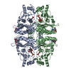
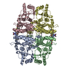
- Assembly
| Deposited unit | 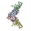
| ||||||||
|---|---|---|---|---|---|---|---|---|---|
| 1 | 
| ||||||||
| 2 | 
| ||||||||
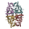
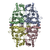
| Unit cell |
|
-Components
| #1: Protein | Mass: 36666.895 Da / Num. of mol.: 4 Source method: isolated from a genetically manipulated source Source: (gene. exp.) Homo sapiens (human) / Tissue: skeletal muscle / Gene: FBP2 / Plasmid: pKK223-3 / Production host:  Escherichia coli (E. coli) / Strain (production host): C100 / References: UniProt: O00757, fructose-bisphosphatase Escherichia coli (E. coli) / Strain (production host): C100 / References: UniProt: O00757, fructose-bisphosphatase#2: Chemical | ChemComp-AMP / Adenosine monophosphate  Mass: 347.221 Da / Num. of mol.: 4 / Source method: obtained synthetically / Formula: C10H14N5O7P / Comment: AMP*YM Mass: 347.221 Da / Num. of mol.: 4 / Source method: obtained synthetically / Formula: C10H14N5O7P / Comment: AMP*YM#3: Water | ChemComp-HOH / | Water Mass: 18.015 Da / Num. of mol.: 664 / Source method: isolated from a natural source / Formula: H2O Mass: 18.015 Da / Num. of mol.: 664 / Source method: isolated from a natural source / Formula: H2O |
|---|
-Experimental details
-Experiment
| Experiment | Method: X-RAY DIFFRACTION |
|---|
- Sample preparation
| Crystal | Density Matthews: 3.06 Å3/Da / Density % sol: 59.86 % |
|---|---|
| Crystal grow | Temperature: 292 K / Method: vapor diffusion, hanging drop / pH: 7 / Details: 1.6M ammonium citrate tribasic / PH range: 7 |
-Data collection
| Diffraction | Mean temperature: 100 K |
|---|---|
| Diffraction source | Source: SYNCHROTRON / Site: BESSY  / Beamline: 14.2 / Wavelength: 0.82657 Å / Beamline: 14.2 / Wavelength: 0.82657 Å |
| Detector | Type: RAYONIX MX-225 / Detector: CCD / Date: Sep 8, 2012 |
| Radiation | Monochromator: Double Crystal Monochromator, Si-111 crystal / Protocol: SINGLE WAVELENGTH / Monochromatic (M) / Laue (L): M / Scattering type: x-ray |
| Radiation wavelength | Wavelength: 0.82657 Å / Relative weight: 1 |
| Reflection | Resolution: 1.85→45.389 Å / Num. all: 156646 / Num. obs: 156646 / % possible obs: 99.2 % / Redundancy: 5.04 % / Biso Wilson estimate: 34.51 Å2 / Rmerge(I) obs: 0.055 / Net I/σ(I): 19.43 |
| Reflection shell | Resolution: 1.85→1.96 Å / Redundancy: 4.98 % / Rmerge(I) obs: 0.693 / Mean I/σ(I) obs: 2.99 / % possible all: 97.4 |
- Processing
| Software |
| |||||||||||||||||||||||||||||||||||||||||||||||||||||||||||||||||||||||||||||||||||||||||||||||||||||||||||||||||||||||||||||||||||||||||||||||||||||||||||||||||||||||||||||||||||||||||||||||||||||||||||||||||||||||||||||||||||||||||||||||||||||||||||||||||||||||||||||||||||||||||||||||||||||||||||||||||||||||||||||||||||||||||||||||||||||||||||||||||||||||||||||||||||||||||||||||||||||||||||||||||||||||||||||||||||||||||
|---|---|---|---|---|---|---|---|---|---|---|---|---|---|---|---|---|---|---|---|---|---|---|---|---|---|---|---|---|---|---|---|---|---|---|---|---|---|---|---|---|---|---|---|---|---|---|---|---|---|---|---|---|---|---|---|---|---|---|---|---|---|---|---|---|---|---|---|---|---|---|---|---|---|---|---|---|---|---|---|---|---|---|---|---|---|---|---|---|---|---|---|---|---|---|---|---|---|---|---|---|---|---|---|---|---|---|---|---|---|---|---|---|---|---|---|---|---|---|---|---|---|---|---|---|---|---|---|---|---|---|---|---|---|---|---|---|---|---|---|---|---|---|---|---|---|---|---|---|---|---|---|---|---|---|---|---|---|---|---|---|---|---|---|---|---|---|---|---|---|---|---|---|---|---|---|---|---|---|---|---|---|---|---|---|---|---|---|---|---|---|---|---|---|---|---|---|---|---|---|---|---|---|---|---|---|---|---|---|---|---|---|---|---|---|---|---|---|---|---|---|---|---|---|---|---|---|---|---|---|---|---|---|---|---|---|---|---|---|---|---|---|---|---|---|---|---|---|---|---|---|---|---|---|---|---|---|---|---|---|---|---|---|---|---|---|---|---|---|---|---|---|---|---|---|---|---|---|---|---|---|---|---|---|---|---|---|---|---|---|---|---|---|---|---|---|---|---|---|---|---|---|---|---|---|---|---|---|---|---|---|---|---|---|---|---|---|---|---|---|---|---|---|---|---|---|---|---|---|---|---|---|---|---|---|---|---|---|---|---|---|---|---|---|---|---|---|---|---|---|---|---|---|---|---|---|---|---|---|---|---|---|---|---|---|---|---|---|---|---|---|---|---|---|---|---|---|---|---|---|---|---|---|---|---|---|---|---|---|---|---|---|---|---|---|---|---|---|---|---|---|---|---|---|---|---|---|---|---|---|---|---|---|---|---|---|---|---|---|---|---|---|---|---|---|---|---|
| Refinement | Method to determine structure: MOLECULAR REPLACEMENT Starting model: 3IFA Resolution: 1.845→45.389 Å / SU ML: 0.19 / Cross valid method: FREE R-VALUE / σ(F): 1.35 / Phase error: 21.21 / Stereochemistry target values: ENGH & HUBER / Details: H atoms were added at riding positions
| |||||||||||||||||||||||||||||||||||||||||||||||||||||||||||||||||||||||||||||||||||||||||||||||||||||||||||||||||||||||||||||||||||||||||||||||||||||||||||||||||||||||||||||||||||||||||||||||||||||||||||||||||||||||||||||||||||||||||||||||||||||||||||||||||||||||||||||||||||||||||||||||||||||||||||||||||||||||||||||||||||||||||||||||||||||||||||||||||||||||||||||||||||||||||||||||||||||||||||||||||||||||||||||||||||||||||
| Solvent computation | Shrinkage radii: 0.9 Å / VDW probe radii: 1.11 Å / Solvent model: FLAT BULK SOLVENT MODEL | |||||||||||||||||||||||||||||||||||||||||||||||||||||||||||||||||||||||||||||||||||||||||||||||||||||||||||||||||||||||||||||||||||||||||||||||||||||||||||||||||||||||||||||||||||||||||||||||||||||||||||||||||||||||||||||||||||||||||||||||||||||||||||||||||||||||||||||||||||||||||||||||||||||||||||||||||||||||||||||||||||||||||||||||||||||||||||||||||||||||||||||||||||||||||||||||||||||||||||||||||||||||||||||||||||||||||
| Refinement step | Cycle: LAST / Resolution: 1.845→45.389 Å
| |||||||||||||||||||||||||||||||||||||||||||||||||||||||||||||||||||||||||||||||||||||||||||||||||||||||||||||||||||||||||||||||||||||||||||||||||||||||||||||||||||||||||||||||||||||||||||||||||||||||||||||||||||||||||||||||||||||||||||||||||||||||||||||||||||||||||||||||||||||||||||||||||||||||||||||||||||||||||||||||||||||||||||||||||||||||||||||||||||||||||||||||||||||||||||||||||||||||||||||||||||||||||||||||||||||||||
| Refine LS restraints |
| |||||||||||||||||||||||||||||||||||||||||||||||||||||||||||||||||||||||||||||||||||||||||||||||||||||||||||||||||||||||||||||||||||||||||||||||||||||||||||||||||||||||||||||||||||||||||||||||||||||||||||||||||||||||||||||||||||||||||||||||||||||||||||||||||||||||||||||||||||||||||||||||||||||||||||||||||||||||||||||||||||||||||||||||||||||||||||||||||||||||||||||||||||||||||||||||||||||||||||||||||||||||||||||||||||||||||
| LS refinement shell |
| |||||||||||||||||||||||||||||||||||||||||||||||||||||||||||||||||||||||||||||||||||||||||||||||||||||||||||||||||||||||||||||||||||||||||||||||||||||||||||||||||||||||||||||||||||||||||||||||||||||||||||||||||||||||||||||||||||||||||||||||||||||||||||||||||||||||||||||||||||||||||||||||||||||||||||||||||||||||||||||||||||||||||||||||||||||||||||||||||||||||||||||||||||||||||||||||||||||||||||||||||||||||||||||||||||||||||
| Refinement TLS params. | Method: refined / Refine-ID: X-RAY DIFFRACTION
| |||||||||||||||||||||||||||||||||||||||||||||||||||||||||||||||||||||||||||||||||||||||||||||||||||||||||||||||||||||||||||||||||||||||||||||||||||||||||||||||||||||||||||||||||||||||||||||||||||||||||||||||||||||||||||||||||||||||||||||||||||||||||||||||||||||||||||||||||||||||||||||||||||||||||||||||||||||||||||||||||||||||||||||||||||||||||||||||||||||||||||||||||||||||||||||||||||||||||||||||||||||||||||||||||||||||||
| Refinement TLS group |
|