 Movie
Movie Controller

[English] 日本語
 Yorodumi
- PDB-5ky2: mouse POFUT1 in complex with O-glucosylated mouse Factor VII EGF1... -
+ Open data

- Basic information
| Entry | Database: PDB / ID: 5ky2 | |||||||||
|---|---|---|---|---|---|---|---|---|---|---|
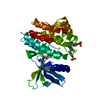
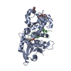
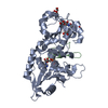
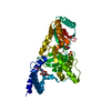
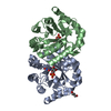
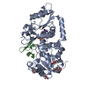
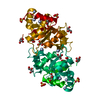
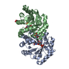
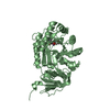
| Title | mouse POFUT1 in complex with O-glucosylated mouse Factor VII EGF1 and GDP | |||||||||
 Components Components |
| |||||||||
 Keywords Keywords |  TRANSFERASE / glycosyltransferase TRANSFERASE / glycosyltransferase | |||||||||
| Function / homology |  Function and homology information Function and homology informationExtrinsic Pathway of Fibrin Clot Formation / Gamma-carboxylation of protein precursors / Transport of gamma-carboxylated protein precursors from the endoplasmic reticulum to the Golgi apparatus / Removal of aminoterminal propeptides from gamma-carboxylated proteins / peptide-O-fucosyltransferase / protein O-linked fucosylation / peptide-O-fucosyltransferase activity / fucosyltransferase activity / regulation of Notch signaling pathway / fucose metabolic process ...Extrinsic Pathway of Fibrin Clot Formation / Gamma-carboxylation of protein precursors / Transport of gamma-carboxylated protein precursors from the endoplasmic reticulum to the Golgi apparatus / Removal of aminoterminal propeptides from gamma-carboxylated proteins / peptide-O-fucosyltransferase / protein O-linked fucosylation / peptide-O-fucosyltransferase activity / fucosyltransferase activity / regulation of Notch signaling pathway / fucose metabolic process / coagulation factor VIIa / response to Thyroid stimulating hormone / response to 2,3,7,8-tetrachlorodibenzodioxine / response to astaxanthin / response to thyrotropin-releasing hormone / response to genistein / serine-type peptidase complex / positive regulation of platelet-derived growth factor receptor signaling pathway / response to vitamin K / response to carbon dioxide / response to thyroxine / protein O-linked glycosylation / response to cholesterol / response to growth hormone / positive regulation of positive chemotaxis / positive regulation of leukocyte chemotaxis / positive regulation of TOR signaling / positive regulation of blood coagulation / animal organ regeneration / somitogenesis / Notch signaling pathway / response to nutrient levels / protein processing / circadian rhythm / response to estrogen / blood coagulation / response to estradiol / nervous system development / heart development / angiogenesis / endopeptidase activity / vesicle / response to hypoxia / serine-type endopeptidase activity / signaling receptor binding / calcium ion binding / endoplasmic reticulum / extracellular space / extracellular region / membraneSimilarity search - Function | |||||||||
| Biological species |  Mus musculus (house mouse) Mus musculus (house mouse) | |||||||||
| Method | X-RAY DIFFRACTION / SYNCHROTRON / MOLECULAR REPLACEMENT / molecular replacement / Resolution: 1.47 Å | |||||||||
 Authors Authors | Li, Z. / Rini, J.M. | |||||||||
| Funding support |  Canada, 1items Canada, 1items
| |||||||||
 Citation Citation | Journal: Nat. Chem. Biol. / Year: 2017 Title: Recognition of EGF-like domains by the Notch-modifying O-fucosyltransferase POFUT1. Authors: Li, Z. / Han, K. / Pak, J.E. / Satkunarajah, M. / Zhou, D. / Rini, J.M. | |||||||||
| History |
|
- Structure visualization
| Structure viewer | Molecule: MolmilJmol/JSmol |
|---|
- Downloads & links
-Download
| PDBx/mmCIF format | 5ky2.cif.gz | 254.9 KB | Display | PDBx/mmCIF format |
|---|---|---|---|---|
| PDB format | pdb5ky2.ent.gz | 211.5 KB | Display | PDB format |
| PDBx/mmJSON format | 5ky2.json.gz | Tree view | PDBx/mmJSON format | |
| Others |  Other downloads Other downloads |
-Validation report
| Arichive directory | https://data.pdbj.org/pub/pdb/validation_reports/ky/5ky2ftp://data.pdbj.org/pub/pdb/validation_reports/ky/5ky2 | HTTPS FTP |
|---|
-Related structure data
| Related structure data |  5kxhC  5kxqC  5ky0C  5ky3C  5ky4C  5ky5C  5ky7C 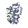 5ky8C  5ky9C  5kx3 C: citing same article ( |
|---|---|
| Similar structure data |


-Links
 PDBj
PDBj


- Assembly
| Deposited unit | 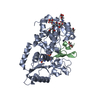
| ||||||||
|---|---|---|---|---|---|---|---|---|---|
| 1 |
| ||||||||
| Unit cell |
|
-Components
-Protein / Protein/peptide , 2 types, 2 molecules AB
| #1: Protein | / Peptide-O-fucosyltransferase 1 / O-FucT-1 Mass: 40457.266 Da / Num. of mol.: 1 Source method: isolated from a genetically manipulated source Source: (gene. exp.) Mus musculus (house mouse) / Gene: Pofut1 / Plasmid: PB-T-PAF / Production host:  Homo sapiens (human) / References: UniProt: Q91ZW2, peptide-O-fucosyltransferase Homo sapiens (human) / References: UniProt: Q91ZW2, peptide-O-fucosyltransferase |
|---|---|
| #2: Protein/peptide | / Serum prothrombin conversion accelerator Mass: 4419.888 Da / Num. of mol.: 1 Source method: isolated from a genetically manipulated source Source: (gene. exp.) Mus musculus (house mouse) / Gene: F7, Cf7 / Production host:  Escherichia coli (E. coli) / References: UniProt: P70375, coagulation factor VIIa Escherichia coli (E. coli) / References: UniProt: P70375, coagulation factor VIIa |
-Sugars , 2 types, 3 molecules 

| #3: Sugar | N-Acetylglucosamine Type: D-saccharide, beta linking / Mass: 221.208 Da / Num. of mol.: 2 Type: D-saccharide, beta linking / Mass: 221.208 Da / Num. of mol.: 2Source method: isolated from a genetically manipulated source Formula: C8H15NO6 #6: Sugar | ChemComp-BGC / | Glucose Type: D-saccharide, beta linking / Mass: 180.156 Da / Num. of mol.: 1 Type: D-saccharide, beta linking / Mass: 180.156 Da / Num. of mol.: 1Source method: isolated from a genetically manipulated source Formula: C6H12O6 |
|---|
-Non-polymers , 3 types, 378 molecules 


| #4: Chemical | ChemComp-GDP / Guanosine diphosphate Type: RNA linking / Mass: 443.201 Da / Num. of mol.: 1 / Source method: obtained synthetically / Formula: C10H15N5O11P2 / Comment: GDP, energy-carrying molecule*YM Type: RNA linking / Mass: 443.201 Da / Num. of mol.: 1 / Source method: obtained synthetically / Formula: C10H15N5O11P2 / Comment: GDP, energy-carrying molecule*YM | ||
|---|---|---|---|
| #5: Chemical | Glycerol Mass: 92.094 Da / Num. of mol.: 3 / Source method: obtained synthetically / Formula: C3H8O3 Mass: 92.094 Da / Num. of mol.: 3 / Source method: obtained synthetically / Formula: C3H8O3#7: Water | ChemComp-HOH / | WaterMass: 18.015 Da / Num. of mol.: 374 / Source method: isolated from a natural source / Formula: H2O |
-Experimental details
-Experiment
| Experiment | Method: X-RAY DIFFRACTION / Number of used crystals: 1 |
|---|
- Sample preparation
| Crystal | Density Matthews: 2.13 Å3/Da / Density % sol: 42.13 % |
|---|---|
| Crystal grow | Temperature: 295 K / Method: vapor diffusion, hanging drop / pH: 8.5 / Details: 20% PEG2000 MME, 50 mM Tris pH 8.5 |
-Data collection
| Diffraction | Mean temperature: 100 K |
|---|---|
| Diffraction source | Source: SYNCHROTRON / Site: CLSI / Beamline: 08ID-1 / Wavelength: 0.97949 Å |
| Detector | Type: RAYONIX MX-300 / Detector: CCD / Date: Apr 1, 2012 |
| Radiation | Protocol: SINGLE WAVELENGTH / Monochromatic (M) / Laue (L): M / Scattering type: x-ray |
| Radiation wavelength | Wavelength: 0.97949 Å / Relative weight: 1 |
| Reflection | Resolution: 1.47→50 Å / Num. obs: 66097 / % possible obs: 100 % / Redundancy: 7.2 % / Biso Wilson estimate: 18 Å2 / CC1/2: 0.994 / Rmerge(I) obs: 0.123 / Net I/σ(I): 8.3 |
| Reflection shell | Resolution: 1.47→1.52 Å / Redundancy: 6.5 % / Rmerge(I) obs: 1.524 / Mean I/σ(I) obs: 1 / CC1/2: 0.466 / % possible all: 100 |
-Phasing
| Phasing | Method: molecular replacement |
|---|
- Processing
| Software |
| |||||||||||||||||||||||||||||||||||||||||||||||||||||||||||||||||||||||||||||||||||||||||||||||||||||||||||||||||||||||||||||||||||||||||||||||||||||||||||||||||||||||||||||||||||||||||||||||||||||||||||||||||||||||||||||||||
|---|---|---|---|---|---|---|---|---|---|---|---|---|---|---|---|---|---|---|---|---|---|---|---|---|---|---|---|---|---|---|---|---|---|---|---|---|---|---|---|---|---|---|---|---|---|---|---|---|---|---|---|---|---|---|---|---|---|---|---|---|---|---|---|---|---|---|---|---|---|---|---|---|---|---|---|---|---|---|---|---|---|---|---|---|---|---|---|---|---|---|---|---|---|---|---|---|---|---|---|---|---|---|---|---|---|---|---|---|---|---|---|---|---|---|---|---|---|---|---|---|---|---|---|---|---|---|---|---|---|---|---|---|---|---|---|---|---|---|---|---|---|---|---|---|---|---|---|---|---|---|---|---|---|---|---|---|---|---|---|---|---|---|---|---|---|---|---|---|---|---|---|---|---|---|---|---|---|---|---|---|---|---|---|---|---|---|---|---|---|---|---|---|---|---|---|---|---|---|---|---|---|---|---|---|---|---|---|---|---|---|---|---|---|---|---|---|---|---|---|---|---|---|---|---|---|---|
| Refinement | Method to determine structure: MOLECULAR REPLACEMENT / Resolution: 1.47→46.935 Å / SU ML: 0.14 / Cross valid method: FREE R-VALUE / σ(F): 1.35 / Phase error: 17.98
| |||||||||||||||||||||||||||||||||||||||||||||||||||||||||||||||||||||||||||||||||||||||||||||||||||||||||||||||||||||||||||||||||||||||||||||||||||||||||||||||||||||||||||||||||||||||||||||||||||||||||||||||||||||||||||||||||
| Solvent computation | Shrinkage radii: 0.9 Å / VDW probe radii: 1.11 Å | |||||||||||||||||||||||||||||||||||||||||||||||||||||||||||||||||||||||||||||||||||||||||||||||||||||||||||||||||||||||||||||||||||||||||||||||||||||||||||||||||||||||||||||||||||||||||||||||||||||||||||||||||||||||||||||||||
| Displacement parameters | Biso max: 120.85 Å2 / Biso mean: 30.2703 Å2 / Biso min: 10.89 Å2 | |||||||||||||||||||||||||||||||||||||||||||||||||||||||||||||||||||||||||||||||||||||||||||||||||||||||||||||||||||||||||||||||||||||||||||||||||||||||||||||||||||||||||||||||||||||||||||||||||||||||||||||||||||||||||||||||||
| Refinement step | Cycle: final / Resolution: 1.47→46.935 Å
| |||||||||||||||||||||||||||||||||||||||||||||||||||||||||||||||||||||||||||||||||||||||||||||||||||||||||||||||||||||||||||||||||||||||||||||||||||||||||||||||||||||||||||||||||||||||||||||||||||||||||||||||||||||||||||||||||
| Refine LS restraints |
| |||||||||||||||||||||||||||||||||||||||||||||||||||||||||||||||||||||||||||||||||||||||||||||||||||||||||||||||||||||||||||||||||||||||||||||||||||||||||||||||||||||||||||||||||||||||||||||||||||||||||||||||||||||||||||||||||
| LS refinement shell | Refine-ID: X-RAY DIFFRACTION / Total num. of bins used: 22
| |||||||||||||||||||||||||||||||||||||||||||||||||||||||||||||||||||||||||||||||||||||||||||||||||||||||||||||||||||||||||||||||||||||||||||||||||||||||||||||||||||||||||||||||||||||||||||||||||||||||||||||||||||||||||||||||||
| Refinement TLS params. | Method: refined / Refine-ID: X-RAY DIFFRACTION
| |||||||||||||||||||||||||||||||||||||||||||||||||||||||||||||||||||||||||||||||||||||||||||||||||||||||||||||||||||||||||||||||||||||||||||||||||||||||||||||||||||||||||||||||||||||||||||||||||||||||||||||||||||||||||||||||||
| Refinement TLS group |
|
