 Movie
Movie Controller

[English] 日本語
 Yorodumi
- PDB-5kr5: Directed Evolution of Transaminases By Ancestral Reconstruction. ... -
+ Open data

- Basic information
| Entry | Database: PDB / ID: 5kr5 | ||||||
|---|---|---|---|---|---|---|---|
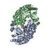
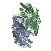
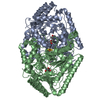
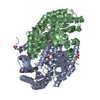
| Title | Directed Evolution of Transaminases By Ancestral Reconstruction. Using Old Proteins for New Chemistries | ||||||
 Components Components | 4-aminobutyrate transaminase | ||||||
 Keywords Keywords | TRANSFERASE / phylogenetics / directed evolution / transaminase / protein engineering | ||||||
| Function / homology | Aspartate Aminotransferase; domain 2 / Type I PLP-dependent aspartate aminotransferase-like (Major domain) / 3-Layer(aba) Sandwich / Alpha Beta / DI(HYDROXYETHYL)ETHER / PYRIDOXAL-5'-PHOSPHATE Function and homology information Function and homology information | ||||||
| Biological species |  Pseudomonas (RNA similarity group I) Pseudomonas (RNA similarity group I) | ||||||
| Method | X-RAY DIFFRACTION / SYNCHROTRON / MOLECULAR REPLACEMENT / Resolution: 2.1 Å | ||||||
 Authors Authors | Wilding, M. / Newman, J. / Peat, T.S. / Scott, C. | ||||||
 Citation Citation | Journal: Green Chemistry / Year: 2017 Title: Reverse engineering: transaminase biocatalyst development using ancestral sequence reconstruction Authors: Wilding, M. / Peat, T.S. / Kalyaanamoorthy, S. / Newman, J. / Scott, C. / Jermiin, L.S. | ||||||
| History |
|
- Structure visualization
| Structure viewer | Molecule: MolmilJmol/JSmol |
|---|
- Downloads & links
-Download
| PDBx/mmCIF format | 5kr5.cif.gz | 195.6 KB | Display | PDBx/mmCIF format |
|---|---|---|---|---|
| PDB format | pdb5kr5.ent.gz | 152.6 KB | Display | PDB format |
| PDBx/mmJSON format | 5kr5.json.gz | Tree view | PDBx/mmJSON format | |
| Others |  Other downloads Other downloads |
-Validation report
| Arichive directory | https://data.pdbj.org/pub/pdb/validation_reports/kr/5kr5ftp://data.pdbj.org/pub/pdb/validation_reports/kr/5kr5 | HTTPS FTP |
|---|
-Related structure data
| Related structure data |  5kqtSC  5kquC  5kqwC  5kr3C  5kr4C  5kr6C S: Starting model for refinement C: citing same article ( |
|---|---|
| Similar structure data |

-Links
 PDBj
PDBj- Assembly
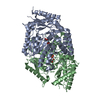
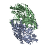
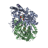
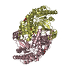
| Deposited unit | 
| ||||||||||||||||||
|---|---|---|---|---|---|---|---|---|---|---|---|---|---|---|---|---|---|---|---|
| 1 |
| ||||||||||||||||||
| Unit cell |
| ||||||||||||||||||
| Noncrystallographic symmetry (NCS) | NCS domain:
NCS domain segments: Component-ID: 0 / Ens-ID: 1 / Beg auth comp-ID: SER / Beg label comp-ID: SER / End auth comp-ID: ALA / End label comp-ID: ALA / Refine code: 0 / Auth seq-ID: 3 - 455 / Label seq-ID: 23 - 475
|
-Components
| #1: Protein | Mass: 52033.895 Da / Num. of mol.: 2 Source method: isolated from a genetically manipulated source Source: (gene. exp.) Pseudomonas (RNA similarity group I) / Plasmid: pET / Production host: Escherichia coli (E. coli) / Strain (production host): BL21 / References: beta-alanine-pyruvate transaminase#2: Chemical | Pyridoxal phosphate  Mass: 247.142 Da / Num. of mol.: 2 Mass: 247.142 Da / Num. of mol.: 2Source method: isolated from a genetically manipulated source Formula: C8H10NO6P / References: beta-alanine-pyruvate transaminase#3: Chemical |   Mass: 40.078 Da / Num. of mol.: 3 / Source method: obtained synthetically / Formula: Ca Mass: 40.078 Da / Num. of mol.: 3 / Source method: obtained synthetically / Formula: Ca#4: Chemical | Diethylene glycol  Mass: 106.120 Da / Num. of mol.: 3 / Source method: obtained synthetically / Formula: C4H10O3 Mass: 106.120 Da / Num. of mol.: 3 / Source method: obtained synthetically / Formula: C4H10O3#5: Water | ChemComp-HOH / | Water Mass: 18.015 Da / Num. of mol.: 358 / Source method: isolated from a natural source / Formula: H2O Mass: 18.015 Da / Num. of mol.: 358 / Source method: isolated from a natural source / Formula: H2O |
|---|
-Experimental details
-Experiment
| Experiment | Method: X-RAY DIFFRACTION / Number of used crystals: 1 |
|---|
- Sample preparation
| Crystal | Density Matthews: 2.42 Å3/Da / Density % sol: 49.22 % |
|---|---|
| Crystal grow | Temperature: 293 K / Method: vapor diffusion, sitting drop Details: 150 plus 150 nL drops with protein at 4 mg/mL and reservoir conditions of 15% PEG 8000, 11 mM calcium acetate, 100 mM Tris pH 7.1. Microcrystals were used as nucleating agents to obtain full size crystals. |
-Data collection
| Diffraction | Mean temperature: 100 K |
|---|---|
| Diffraction source | Source: SYNCHROTRON / Site: Australian Synchrotron  / Beamline: MX2 / Wavelength: 0.9537 Å / Beamline: MX2 / Wavelength: 0.9537 Å |
| Detector | Type: ADSC QUANTUM 315r / Detector: CCD / Date: Mar 1, 2014 |
| Radiation | Protocol: SINGLE WAVELENGTH / Monochromatic (M) / Laue (L): M / Scattering type: x-ray |
| Radiation wavelength | Wavelength: 0.9537 Å / Relative weight: 1 |
| Reflection | Resolution: 2.1→48.6 Å / Num. obs: 59261 / % possible obs: 99.6 % / Redundancy: 7.3 % / CC1/2: 0.994 / Rmerge(I) obs: 0.17 / Net I/σ(I): 9.5 |
| Reflection shell | Resolution: 2.1→2.15 Å / Redundancy: 7.3 % / Rmerge(I) obs: 0.899 / Mean I/σ(I) obs: 2.2 / CC1/2: 0.67 / % possible all: 97.6 |
- Processing
| Software |
| ||||||||||||||||||||||||||||||||||||||||||||||||||||||||||||||||||||||||||||||||||||||||||||||||||||||||||||||||||||||||||||||||||||||||||||||||||||||||||||||||||||||||||||||||||||||
|---|---|---|---|---|---|---|---|---|---|---|---|---|---|---|---|---|---|---|---|---|---|---|---|---|---|---|---|---|---|---|---|---|---|---|---|---|---|---|---|---|---|---|---|---|---|---|---|---|---|---|---|---|---|---|---|---|---|---|---|---|---|---|---|---|---|---|---|---|---|---|---|---|---|---|---|---|---|---|---|---|---|---|---|---|---|---|---|---|---|---|---|---|---|---|---|---|---|---|---|---|---|---|---|---|---|---|---|---|---|---|---|---|---|---|---|---|---|---|---|---|---|---|---|---|---|---|---|---|---|---|---|---|---|---|---|---|---|---|---|---|---|---|---|---|---|---|---|---|---|---|---|---|---|---|---|---|---|---|---|---|---|---|---|---|---|---|---|---|---|---|---|---|---|---|---|---|---|---|---|---|---|---|---|
| Refinement | Method to determine structure: MOLECULAR REPLACEMENT Starting model: 5kqt Resolution: 2.1→48.6 Å / Cor.coef. Fo:Fc: 0.938 / Cor.coef. Fo:Fc free: 0.908 / SU B: 6.161 / SU ML: 0.155 / Cross valid method: THROUGHOUT / ESU R: 0.23 / ESU R Free: 0.186 / Details: HYDROGENS HAVE BEEN ADDED IN THE RIDING POSITIONS
| ||||||||||||||||||||||||||||||||||||||||||||||||||||||||||||||||||||||||||||||||||||||||||||||||||||||||||||||||||||||||||||||||||||||||||||||||||||||||||||||||||||||||||||||||||||||
| Solvent computation | Ion probe radii: 0.8 Å / Shrinkage radii: 0.8 Å / VDW probe radii: 1.2 Å | ||||||||||||||||||||||||||||||||||||||||||||||||||||||||||||||||||||||||||||||||||||||||||||||||||||||||||||||||||||||||||||||||||||||||||||||||||||||||||||||||||||||||||||||||||||||
| Displacement parameters | Biso mean: 29.309 Å2
| ||||||||||||||||||||||||||||||||||||||||||||||||||||||||||||||||||||||||||||||||||||||||||||||||||||||||||||||||||||||||||||||||||||||||||||||||||||||||||||||||||||||||||||||||||||||
| Refinement step | Cycle: 1 / Resolution: 2.1→48.6 Å
| ||||||||||||||||||||||||||||||||||||||||||||||||||||||||||||||||||||||||||||||||||||||||||||||||||||||||||||||||||||||||||||||||||||||||||||||||||||||||||||||||||||||||||||||||||||||
| Refine LS restraints |
|