 Movie
Movie Controller

[English] 日本語
 Yorodumi
- PDB-5klh: Crystal Structure of Trypanosoma brucei Procyclic Specific Surfac... -
+ Open data

- Basic information
| Entry | Database: PDB / ID: 5klh | ||||||
|---|---|---|---|---|---|---|---|
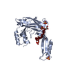
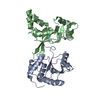
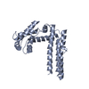
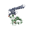
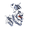
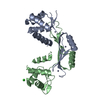
| Title | Crystal Structure of Trypanosoma brucei Procyclic Specific Surface Antigen-2 | ||||||
 Components Components | Surface glycoprotein | ||||||
 Keywords Keywords |  MEMBRANE PROTEIN / Bi-lobed architecture / surface protein / putative sensor MEMBRANE PROTEIN / Bi-lobed architecture / surface protein / putative sensor | ||||||
| Function / homology | membrane => GO:0016020 / Surface glycoprotein Function and homology information Function and homology information | ||||||
| Biological species |  Trypanosoma brucei (eukaryote) Trypanosoma brucei (eukaryote) | ||||||
| Method | X-RAY DIFFRACTION / SYNCHROTRON / SAD / Resolution: 1.646 Å | ||||||
 Authors Authors | Ramaswamy, R. / Parker, M.L. / Boulanger, M.J. | ||||||
| Funding support |  Canada, 1items Canada, 1items
| ||||||
 Citation Citation | Journal: Protein Sci. / Year: 2016 Title: Structural characterization reveals a novel bilobed architecture for the ectodomains of insect stage expressed Trypanosoma brucei PSSA-2 and Trypanosoma congolense ISA. Authors: Ramaswamy, R. / Goomeshi Nobary, S. / Eyford, B.A. / Pearson, T.W. / Boulanger, M.J. | ||||||
| History |
|
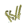
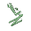
- Structure visualization
| Structure viewer | Molecule: MolmilJmol/JSmol |
|---|
- Downloads & links
-Download
| PDBx/mmCIF format | 5klh.cif.gz | 106 KB | Display | PDBx/mmCIF format |
|---|---|---|---|---|
| PDB format | pdb5klh.ent.gz | 84 KB | Display | PDB format |
| PDBx/mmJSON format | 5klh.json.gz | Tree view | PDBx/mmJSON format | |
| Others |  Other downloads Other downloads |
-Validation report
| Arichive directory | https://data.pdbj.org/pub/pdb/validation_reports/kl/5klhftp://data.pdbj.org/pub/pdb/validation_reports/kl/5klh | HTTPS FTP |
|---|
-Related structure data


-Links
 PDBj
PDBj- Assembly
| Deposited unit | 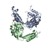
| ||||||||
|---|---|---|---|---|---|---|---|---|---|
| 1 | 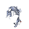
| ||||||||
| 2 | 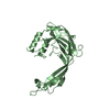
| ||||||||
| Unit cell |
|
-Components
| #1: Protein | Mass: 25393.121 Da / Num. of mol.: 2 / Fragment: residues 40-262 Source method: isolated from a genetically manipulated source Source: (gene. exp.) Trypanosoma brucei (eukaryote) / Plasmid: pET28(a) / Production host:  Escherichia coli (E. coli) / References: UniProt: Q26806 Escherichia coli (E. coli) / References: UniProt: Q26806#2: Chemical | ChemComp-SO4 / | Sulfate  Mass: 96.063 Da / Num. of mol.: 1 / Source method: obtained synthetically / Formula: SO4 Mass: 96.063 Da / Num. of mol.: 1 / Source method: obtained synthetically / Formula: SO4#3: Water | ChemComp-HOH / | Water Mass: 18.015 Da / Num. of mol.: 462 / Source method: isolated from a natural source / Formula: H2O Mass: 18.015 Da / Num. of mol.: 462 / Source method: isolated from a natural source / Formula: H2O |
|---|
-Experimental details
-Experiment
| Experiment | Method: X-RAY DIFFRACTION / Number of used crystals: 1 |
|---|
- Sample preparation
| Crystal | Density Matthews: 2.12 Å3/Da / Density % sol: 41.98 % |
|---|---|
| Crystal grow | Temperature: 291 K / Method: vapor diffusion, sitting drop / pH: 6.5 Details: 0.2 M (NH4)2SO4, 0.1 M Bis-tris pH 6.5, and 25% PEG 3350 |
-Data collection
| Diffraction | Mean temperature: 100 K |
|---|---|
| Diffraction source | Source: SYNCHROTRON / Site: SSRL  / Beamline: BL9-2 / Wavelength: 0.9795 Å / Beamline: BL9-2 / Wavelength: 0.9795 Å |
| Detector | Type: DECTRIS PILATUS 6M / Detector: PIXEL / Date: Nov 12, 2014 |
| Radiation | Protocol: SINGLE WAVELENGTH / Monochromatic (M) / Laue (L): M / Scattering type: x-ray |
| Radiation wavelength | Wavelength: 0.9795 Å / Relative weight: 1 |
| Reflection | Resolution: 1.646→56.95 Å / Num. obs: 45400 / % possible obs: 92.3 % / Redundancy: 3.8 % / Biso Wilson estimate: 21.2 Å2 / CC1/2: 0.995 / Rmerge(I) obs: 0.066 / Net I/σ(I): 10.3 |
- Processing
| Software |
| |||||||||||||||||||||||||||||||||||||||||||||||||||||||||||||||||||||||||||||||||||||||||||||||||||||||||||||||||||||||
|---|---|---|---|---|---|---|---|---|---|---|---|---|---|---|---|---|---|---|---|---|---|---|---|---|---|---|---|---|---|---|---|---|---|---|---|---|---|---|---|---|---|---|---|---|---|---|---|---|---|---|---|---|---|---|---|---|---|---|---|---|---|---|---|---|---|---|---|---|---|---|---|---|---|---|---|---|---|---|---|---|---|---|---|---|---|---|---|---|---|---|---|---|---|---|---|---|---|---|---|---|---|---|---|---|---|---|---|---|---|---|---|---|---|---|---|---|---|---|---|---|
| Refinement | Method to determine structure: SAD / Resolution: 1.646→47.122 Å / SU ML: 0.19 / Cross valid method: FREE R-VALUE / σ(F): 1.98 / Phase error: 22.36
| |||||||||||||||||||||||||||||||||||||||||||||||||||||||||||||||||||||||||||||||||||||||||||||||||||||||||||||||||||||||
| Solvent computation | Shrinkage radii: 0.9 Å / VDW probe radii: 1.11 Å | |||||||||||||||||||||||||||||||||||||||||||||||||||||||||||||||||||||||||||||||||||||||||||||||||||||||||||||||||||||||
| Displacement parameters | Biso max: 66.79 Å2 / Biso mean: 21.38 Å2 / Biso min: 6.62 Å2 | |||||||||||||||||||||||||||||||||||||||||||||||||||||||||||||||||||||||||||||||||||||||||||||||||||||||||||||||||||||||
| Refinement step | Cycle: final / Resolution: 1.646→47.122 Å
| |||||||||||||||||||||||||||||||||||||||||||||||||||||||||||||||||||||||||||||||||||||||||||||||||||||||||||||||||||||||
| Refine LS restraints |
| |||||||||||||||||||||||||||||||||||||||||||||||||||||||||||||||||||||||||||||||||||||||||||||||||||||||||||||||||||||||
| LS refinement shell | Refine-ID: X-RAY DIFFRACTION / Total num. of bins used: 16
|
