 Movie
Movie Controller

[English] 日本語
 Yorodumi
- PDB-5k8y: Structure of the Mus musclus Langerin carbohydrate recognition domain -
+ Open data

- Basic information
| Entry | Database: PDB / ID: 5k8y | ||||||
|---|---|---|---|---|---|---|---|
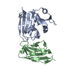
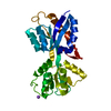
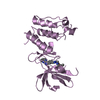
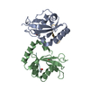
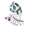
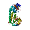
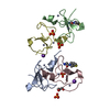
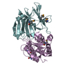
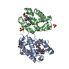
| Title | Structure of the Mus musclus Langerin carbohydrate recognition domain | ||||||
 Components Components | C-type lectin domain family 4 member K | ||||||
 Keywords Keywords |  IMMUNE SYSTEM / C-TYPE LECTIN / GLYCOPROTEIN / CARBOHYDRATE BINDING PROTEIN / CALCIUM BINDING / CRD Domain / LECTIN IMMUNE SYSTEM / C-TYPE LECTIN / GLYCOPROTEIN / CARBOHYDRATE BINDING PROTEIN / CALCIUM BINDING / CRD Domain / LECTIN | ||||||
| Function / homology |  Function and homology information Function and homology informationCross-presentation of soluble exogenous antigens (endosomes) / carbohydrate binding / defense response to virus / external side of plasma membrane / plasma membraneSimilarity search - Function | ||||||
| Biological species |  Mus musculus (house mouse) Mus musculus (house mouse) | ||||||
| Method | X-RAY DIFFRACTION / SYNCHROTRON / MOLECULAR REPLACEMENT / Resolution: 2.4 Å | ||||||
 Authors Authors | Loll, B. / Aretz, J. / Rademacher, C. / Wahl, M.C. | ||||||
 Citation Citation | Journal: J. Biol. Chem. / Year: 2017 Title: Bacterial Polysaccharide Specificity of the Pattern Recognition Receptor Langerin Is Highly Species-dependent. Authors: Hanske, J. / Schulze, J. / Aretz, J. / McBride, R. / Loll, B. / Schmidt, H. / Knirel, Y. / Rabsch, W. / Wahl, M.C. / Paulson, J.C. / Rademacher, C. | ||||||
| History |
|
- Structure visualization
| Structure viewer | Molecule: MolmilJmol/JSmol |
|---|
- Downloads & links
-Download
| PDBx/mmCIF format | 5k8y.cif.gz | 74.1 KB | Display | PDBx/mmCIF format |
|---|---|---|---|---|
| PDB format | pdb5k8y.ent.gz | 54.4 KB | Display | PDB format |
| PDBx/mmJSON format | 5k8y.json.gz | Tree view | PDBx/mmJSON format | |
| Others |  Other downloads Other downloads |
-Validation report
| Arichive directory | https://data.pdbj.org/pub/pdb/validation_reports/k8/5k8yftp://data.pdbj.org/pub/pdb/validation_reports/k8/5k8y | HTTPS FTP |
|---|
-Related structure data
| Related structure data | 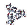 5m62C  3kqgS S: Starting model for refinement C: citing same article ( |
|---|---|
| Similar structure data |

-Links
 PDBj
PDBj


- Assembly
| Deposited unit | 
| |||||||||
|---|---|---|---|---|---|---|---|---|---|---|
| 1 |
| |||||||||
| Unit cell |
| |||||||||
| Components on special symmetry positions |
|
-Components
| #1: Protein | Mass: 17533.578 Da / Num. of mol.: 2 / Fragment: UNP residues 189-326 Source method: isolated from a genetically manipulated source Details: Ala2 and Gly3 are due to cloning / Source: (gene. exp.) Mus musculus (house mouse) / Gene: Cd207, Clec4k / Plasmid: pUC19 / Production host:  Escherichia coli BL21(DE3) (bacteria) / References: UniProt: Q8VBX4 Escherichia coli BL21(DE3) (bacteria) / References: UniProt: Q8VBX4#2: Chemical |   Mass: 40.078 Da / Num. of mol.: 2 / Source method: obtained synthetically / Formula: Ca Mass: 40.078 Da / Num. of mol.: 2 / Source method: obtained synthetically / Formula: Ca#3: Chemical | Polyethylene glycol  Mass: 282.331 Da / Num. of mol.: 3 / Source method: obtained synthetically / Formula: C12H26O7 / Comment: precipitant*YM Mass: 282.331 Da / Num. of mol.: 3 / Source method: obtained synthetically / Formula: C12H26O7 / Comment: precipitant*YM#4: Chemical | ChemComp-GOL / | Glycerol  Mass: 92.094 Da / Num. of mol.: 1 / Source method: obtained synthetically / Formula: C3H8O3 Mass: 92.094 Da / Num. of mol.: 1 / Source method: obtained synthetically / Formula: C3H8O3#5: Water | ChemComp-HOH / | Water Mass: 18.015 Da / Num. of mol.: 80 / Source method: isolated from a natural source / Formula: H2O Mass: 18.015 Da / Num. of mol.: 80 / Source method: isolated from a natural source / Formula: H2O |
|---|
-Experimental details
-Experiment
| Experiment | Method: X-RAY DIFFRACTION / Number of used crystals: 1 |
|---|
- Sample preparation
| Crystal | Density Matthews: 3.5 Å3/Da / Density % sol: 64.83 % |
|---|---|
| Crystal grow | Temperature: 291 K / Method: vapor diffusion, sitting drop / pH: 6 Details: 0.1 M MES, pH 6.0, 30% (v/v) polyethylene glycol 600, 5% (w/v) polyethylene glycol 1000, and 10% (v/v) glycerol |
-Data collection
| Diffraction | Mean temperature: 100 K | |||||||||||||||
|---|---|---|---|---|---|---|---|---|---|---|---|---|---|---|---|---|
| Diffraction source | Source: SYNCHROTRON / Site: BESSY  / Beamline: 14.1 / Wavelength: 0.91841 Å / Beamline: 14.1 / Wavelength: 0.91841 Å | |||||||||||||||
| Detector | Type: DECTRIS PILATUS3 6M / Detector: PIXEL / Date: Mar 4, 2016 / Details: SAGITALLY FOCUSED SI(111) | |||||||||||||||
| Radiation | Protocol: SINGLE WAVELENGTH / Monochromatic (M) / Laue (L): M / Scattering type: x-ray | |||||||||||||||
| Radiation wavelength | Wavelength: 0.91841 Å / Relative weight: 1 | |||||||||||||||
| Reflection twin |
| |||||||||||||||
| Reflection | Resolution: 2.4→50 Å / Num. obs: 19343 / % possible obs: 100 % / Redundancy: 20 % / CC1/2: 0.958 / Rmerge(I) obs: 0.78 / Net I/σ(I): 4.8 | |||||||||||||||
| Reflection shell | Resolution: 2.4→2.54 Å / Redundancy: 19.5 % / Mean I/σ(I) obs: 2.1 / % possible all: 99.9 |
- Processing
| Software |
| ||||||||||||||||||||||||||||||||||||||||||||||||||||||||||||||||||||||||||||||||||||||||||||||||||||||||||||||||||||||||||||||||||||||||||||||||||||||||||||||||||||||||||||||||||||||
|---|---|---|---|---|---|---|---|---|---|---|---|---|---|---|---|---|---|---|---|---|---|---|---|---|---|---|---|---|---|---|---|---|---|---|---|---|---|---|---|---|---|---|---|---|---|---|---|---|---|---|---|---|---|---|---|---|---|---|---|---|---|---|---|---|---|---|---|---|---|---|---|---|---|---|---|---|---|---|---|---|---|---|---|---|---|---|---|---|---|---|---|---|---|---|---|---|---|---|---|---|---|---|---|---|---|---|---|---|---|---|---|---|---|---|---|---|---|---|---|---|---|---|---|---|---|---|---|---|---|---|---|---|---|---|---|---|---|---|---|---|---|---|---|---|---|---|---|---|---|---|---|---|---|---|---|---|---|---|---|---|---|---|---|---|---|---|---|---|---|---|---|---|---|---|---|---|---|---|---|---|---|---|---|
| Refinement | Method to determine structure: MOLECULAR REPLACEMENT Starting model: 3KQG Resolution: 2.4→45.32 Å / Cor.coef. Fo:Fc: 0.916 / Cor.coef. Fo:Fc free: 0.894 / SU B: 3.138 / SU ML: 0.077 / Cross valid method: THROUGHOUT / ESU R: 0.052 / ESU R Free: 0.043
| ||||||||||||||||||||||||||||||||||||||||||||||||||||||||||||||||||||||||||||||||||||||||||||||||||||||||||||||||||||||||||||||||||||||||||||||||||||||||||||||||||||||||||||||||||||||
| Solvent computation | Ion probe radii: 0.8 Å / Shrinkage radii: 0.8 Å / VDW probe radii: 1.2 Å | ||||||||||||||||||||||||||||||||||||||||||||||||||||||||||||||||||||||||||||||||||||||||||||||||||||||||||||||||||||||||||||||||||||||||||||||||||||||||||||||||||||||||||||||||||||||
| Displacement parameters | Biso mean: 52.106 Å2
| ||||||||||||||||||||||||||||||||||||||||||||||||||||||||||||||||||||||||||||||||||||||||||||||||||||||||||||||||||||||||||||||||||||||||||||||||||||||||||||||||||||||||||||||||||||||
| Refinement step | Cycle: LAST / Resolution: 2.4→45.32 Å
| ||||||||||||||||||||||||||||||||||||||||||||||||||||||||||||||||||||||||||||||||||||||||||||||||||||||||||||||||||||||||||||||||||||||||||||||||||||||||||||||||||||||||||||||||||||||
| Refine LS restraints |
|