 Movie
Movie Controller

[English] 日本語
 Yorodumi
- PDB-5k31: Crystal structure of Human fibrillar procollagen type I C-propept... -
+ Open data

- Basic information
| Entry | Database: PDB / ID: 5k31 | ||||||
|---|---|---|---|---|---|---|---|
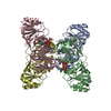
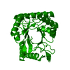
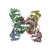
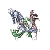
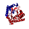
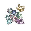
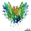
| Title | Crystal structure of Human fibrillar procollagen type I C-propeptide Homo-trimer | ||||||
 Components Components | Collagen alpha-1(I) chain | ||||||
 Keywords Keywords |  STRUCTURAL PROTEIN / FIBRILLAR COLLAGEN / EXTRACELLULAR MATRIX / FIBROSIS STRUCTURAL PROTEIN / FIBRILLAR COLLAGEN / EXTRACELLULAR MATRIX / FIBROSIS | ||||||
| Function / homology |  Function and homology information Function and homology informationcellular response to fluoride / collagen type I trimer / tooth mineralization / cellular response to vitamin E / bone trabecula formation / Anchoring fibril formation / Crosslinking of collagen fibrils / collagen biosynthetic process / Collagen chain trimerization / extracellular matrix structural constituent conferring tensile strength ...cellular response to fluoride / collagen type I trimer / tooth mineralization / cellular response to vitamin E / bone trabecula formation / Anchoring fibril formation / Crosslinking of collagen fibrils / collagen biosynthetic process / Collagen chain trimerization / extracellular matrix structural constituent conferring tensile strength / platelet-derived growth factor binding / intramembranous ossification / Extracellular matrix organization / embryonic skeletal system development / cartilage development involved in endochondral bone morphogenesis / Collagen biosynthesis and modifying enzymes / skin morphogenesis / collagen-activated tyrosine kinase receptor signaling pathway / Platelet Adhesion to exposed collagen / endochondral ossification / cellular response to fibroblast growth factor stimulus / collagen fibril organization / negative regulation of cell-substrate adhesion / face morphogenesis / response to steroid hormone / Scavenging by Class A Receptors / skin development / MET activates PTK2 signaling / Assembly of collagen fibrils and other multimeric structures / Syndecan interactions / GP1b-IX-V activation signalling / blood vessel development / RUNX2 regulates osteoblast differentiation / Platelet Aggregation (Plug Formation) / Collagen degradation / protein localization to nucleus / Non-integrin membrane-ECM interactions / ECM proteoglycans / response to hyperoxia / positive regulation of epithelial to mesenchymal transition / Integrin cell surface interactions / response to mechanical stimulus / cellular response to retinoic acid / response to cAMP / cellular response to epidermal growth factor stimulus / GPVI-mediated activation cascade / cellular response to transforming growth factor beta stimulus / visual perception / extracellular matrix organization / ossification / secretory granule / skeletal system development / cellular response to glucose stimulus / cellular response to amino acid stimulus / Cell surface interactions at the vascular wall / sensory perception of sound / response to insulin / response to hydrogen peroxide / osteoblast differentiation / cellular response to mechanical stimulus / positive regulation of canonical Wnt signaling pathway / Immunoregulatory interactions between a Lymphoid and a non-Lymphoid cell / protein transport / response to estradiol / cellular response to tumor necrosis factor / collagen-containing extracellular matrix / protease binding / positive regulation of cell migration / response to xenobiotic stimulus / endoplasmic reticulum lumen / positive regulation of DNA-templated transcription / extracellular space / extracellular region / identical protein binding / metal ion binding / cytoplasmSimilarity search - Function | ||||||
| Biological species |  Homo sapiens (human) Homo sapiens (human) | ||||||
| Method | X-RAY DIFFRACTION / SYNCHROTRON / MOLECULAR REPLACEMENT / Resolution: 2.2 Å | ||||||
 Authors Authors | Sharma, U. / Hulmes, D.J.S. / Aghajari, N. | ||||||
| Funding support |  France, 1items France, 1items
| ||||||
 Citation Citation | Journal: Nat Commun / Year: 2017 Title: Structural basis of homo- and heterotrimerization of collagen I. Authors: Sharma, U. / Carrique, L. / Vadon-Le Goff, S. / Mariano, N. / Georges, R.N. / Delolme, F. / Koivunen, P. / Myllyharju, J. / Moali, C. / Aghajari, N. / Hulmes, D.J. | ||||||
| History |
|
- Structure visualization
| Structure viewer | Molecule: MolmilJmol/JSmol |
|---|
- Downloads & links
-Download
| PDBx/mmCIF format | 5k31.cif.gz | 568.3 KB | Display | PDBx/mmCIF format |
|---|---|---|---|---|
| PDB format | pdb5k31.ent.gz | 469.1 KB | Display | PDB format |
| PDBx/mmJSON format | 5k31.json.gz | Tree view | PDBx/mmJSON format | |
| Others |  Other downloads Other downloads |
-Validation report
| Arichive directory | https://data.pdbj.org/pub/pdb/validation_reports/k3/5k31ftp://data.pdbj.org/pub/pdb/validation_reports/k3/5k31 | HTTPS FTP |
|---|
-Related structure data
| Related structure data |  2aejS S: Starting model for refinement |
|---|---|
| Similar structure data |

-Links
 PDBj
PDBj


- Assembly
| Deposited unit | 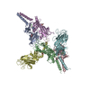
| ||||||||
|---|---|---|---|---|---|---|---|---|---|
| 1 | 
| ||||||||
| 2 | 
| ||||||||
| 3 | 
| ||||||||
| Unit cell |
|
-Components
| #1: Protein | Mass: 28687.947 Da / Num. of mol.: 6 Source method: isolated from a genetically manipulated source Source: (gene. exp.) Homo sapiens (human) / Gene: COL1A1 / Plasmid: PHLSEC / Cell (production host): HEK 293T / Production host: Homo sapiens (human) / References: UniProt: P02452#2: Chemical | ChemComp-CA /   Mass: 40.078 Da / Num. of mol.: 6 / Source method: obtained synthetically / Formula: Ca Mass: 40.078 Da / Num. of mol.: 6 / Source method: obtained synthetically / Formula: Ca#3: Chemical | ChemComp-GOL / Glycerol  Mass: 92.094 Da / Num. of mol.: 5 / Source method: obtained synthetically / Formula: C3H8O3 Mass: 92.094 Da / Num. of mol.: 5 / Source method: obtained synthetically / Formula: C3H8O3#4: Chemical | Chloride  Mass: 35.453 Da / Num. of mol.: 3 / Source method: obtained synthetically / Formula: Cl Mass: 35.453 Da / Num. of mol.: 3 / Source method: obtained synthetically / Formula: Cl#5: Water | ChemComp-HOH / | Water Mass: 18.015 Da / Num. of mol.: 420 / Source method: isolated from a natural source / Formula: H2O Mass: 18.015 Da / Num. of mol.: 420 / Source method: isolated from a natural source / Formula: H2O |
|---|
-Experimental details
-Experiment
| Experiment | Method: X-RAY DIFFRACTION / Number of used crystals: 1 |
|---|
- Sample preparation
| Crystal | Density Matthews: 3.37 Å3/Da / Density % sol: 63.55 % |
|---|---|
| Crystal grow | Temperature: 292 K / Method: vapor diffusion, hanging drop / Details: 18 % PEG 4000 and 0.1 M Tris pH 8.0 |
-Data collection
| Diffraction | Mean temperature: 100 K |
|---|---|
| Diffraction source | Source: SYNCHROTRON / Site: SLS  / Beamline: X06DA / Wavelength: 1 Å / Beamline: X06DA / Wavelength: 1 Å |
| Detector | Type: DECTRIS PILATUS 2M-F / Detector: PIXEL / Date: Oct 13, 2013 |
| Radiation | Protocol: SINGLE WAVELENGTH / Monochromatic (M) / Laue (L): M / Scattering type: x-ray |
| Radiation wavelength | Wavelength: 1 Å / Relative weight: 1 |
| Reflection | Resolution: 2.2→47.07 Å / Num. obs: 115206 / % possible obs: 99.8 % / Redundancy: 3.4 % / Rmerge(I) obs: 0.078 / Net I/σ(I): 7.7 |
| Reflection shell | Highest resolution: 2.2 Å |
- Processing
| Software |
| ||||||||||||||||||||||||||||||||||||||||||||||||||||||||||||||||||||||||||||||||||||||||||||||||||||||||||||||||||||||||||||||||||||||||||||||||||||||||||||||||||||||||||||||||||||||
|---|---|---|---|---|---|---|---|---|---|---|---|---|---|---|---|---|---|---|---|---|---|---|---|---|---|---|---|---|---|---|---|---|---|---|---|---|---|---|---|---|---|---|---|---|---|---|---|---|---|---|---|---|---|---|---|---|---|---|---|---|---|---|---|---|---|---|---|---|---|---|---|---|---|---|---|---|---|---|---|---|---|---|---|---|---|---|---|---|---|---|---|---|---|---|---|---|---|---|---|---|---|---|---|---|---|---|---|---|---|---|---|---|---|---|---|---|---|---|---|---|---|---|---|---|---|---|---|---|---|---|---|---|---|---|---|---|---|---|---|---|---|---|---|---|---|---|---|---|---|---|---|---|---|---|---|---|---|---|---|---|---|---|---|---|---|---|---|---|---|---|---|---|---|---|---|---|---|---|---|---|---|---|---|
| Refinement | Method to determine structure: MOLECULAR REPLACEMENT Starting model: 2AEJ Resolution: 2.2→47.07 Å / Cor.coef. Fo:Fc: 0.947 / Cor.coef. Fo:Fc free: 0.923 / SU B: 11.206 / SU ML: 0.126 / Cross valid method: THROUGHOUT / ESU R: 0.187 / ESU R Free: 0.173 / Details: HYDROGENS HAVE BEEN ADDED IN THE RIDING POSITIONS
| ||||||||||||||||||||||||||||||||||||||||||||||||||||||||||||||||||||||||||||||||||||||||||||||||||||||||||||||||||||||||||||||||||||||||||||||||||||||||||||||||||||||||||||||||||||||
| Solvent computation | Ion probe radii: 0.8 Å / Shrinkage radii: 0.8 Å / VDW probe radii: 1.2 Å | ||||||||||||||||||||||||||||||||||||||||||||||||||||||||||||||||||||||||||||||||||||||||||||||||||||||||||||||||||||||||||||||||||||||||||||||||||||||||||||||||||||||||||||||||||||||
| Displacement parameters | Biso mean: 41.469 Å2
| ||||||||||||||||||||||||||||||||||||||||||||||||||||||||||||||||||||||||||||||||||||||||||||||||||||||||||||||||||||||||||||||||||||||||||||||||||||||||||||||||||||||||||||||||||||||
| Refinement step | Cycle: 1 / Resolution: 2.2→47.07 Å
| ||||||||||||||||||||||||||||||||||||||||||||||||||||||||||||||||||||||||||||||||||||||||||||||||||||||||||||||||||||||||||||||||||||||||||||||||||||||||||||||||||||||||||||||||||||||
| Refine LS restraints |
|