OXIDOREDUCTASE / Rossmann fold / NAD / GAPDH / Glycolysis
Function / homology
Function and homology information
Oxidoreductases; Acting on the aldehyde or oxo group of donors; With NAD+ or NADP+ as acceptor / glyceraldehyde-3-phosphate dehydrogenase (NAD+) (phosphorylating) activity / glycolytic process / glucose metabolic process / NAD binding / NADP binding / identical protein binding / metal ion binding / cytoplasm Similarity search - Function
Glyceraldehyde-3-phosphate dehydrogenase, type I / Glyceraldehyde 3-phosphate dehydrogenase, active site / Glyceraldehyde 3-phosphate dehydrogenase active site. / Glyceraldehyde 3-phosphate dehydrogenase, NAD binding domain / Glyceraldehyde 3-phosphate dehydrogenase, NAD(P) binding domain / Glyceraldehyde 3-phosphate dehydrogenase, catalytic domain / Glyceraldehyde/Erythrose phosphate dehydrogenase family / Glyceraldehyde 3-phosphate dehydrogenase, C-terminal domain / Glyceraldehyde 3-phosphate dehydrogenase, NAD binding domain / Dihydrodipicolinate Reductase; domain 2 ...Glyceraldehyde-3-phosphate dehydrogenase, type I / Glyceraldehyde 3-phosphate dehydrogenase, active site / Glyceraldehyde 3-phosphate dehydrogenase active site. / Glyceraldehyde 3-phosphate dehydrogenase, NAD binding domain / Glyceraldehyde 3-phosphate dehydrogenase, NAD(P) binding domain / Glyceraldehyde 3-phosphate dehydrogenase, catalytic domain / Glyceraldehyde/Erythrose phosphate dehydrogenase family / Glyceraldehyde 3-phosphate dehydrogenase, C-terminal domain / Glyceraldehyde 3-phosphate dehydrogenase, NAD binding domain / Dihydrodipicolinate Reductase; domain 2 / Dihydrodipicolinate Reductase; domain 2 / NAD(P)-binding Rossmann-like Domain / NAD(P)-binding domain superfamily / Rossmann fold / 2-Layer Sandwich / 3-Layer(aba) Sandwich / Alpha Beta Similarity search - Domain/homology
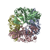
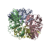
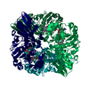
Mass: 38214.867 Da / Num. of mol.: 4 Source method: isolated from a genetically manipulated source Source: (gene. exp.) Streptococcus agalactiae (bacteria) Gene: gap, gapA, EN72_09590, ERS039640_00200, ERS046921_00390, RDF_1710 Plasmid: PET15B / Cell line (production host): Rosetta(DE3)pLysS / Production host: Escherichia coli (E. coli) References: UniProt: Q9ALW2, Oxidoreductases; Acting on the aldehyde or oxo group of donors; With NAD+ or NADP+ as acceptor
 Movie
Movie Controller

 Yorodumi
 Open data
 Basic information
 Components
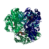
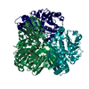
 Glyceraldehyde 3-phosphate dehydrogenase
  Keywords
 Function and homology information
 Authors
 Citation
 Structure visualization
 Downloads & links
 Other downloads
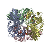


 PDBj
PDBj
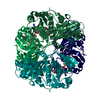
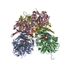
 Assembly
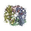

 Mass: 663.425 Da / Num. of mol.: 4 / Source method: obtained synthetically / Formula: C21H27N7O14P2 / Comment: NAD*YM
 Mass: 24.305 Da / Num. of mol.: 4 / Source method: obtained synthetically / Formula: Mg
 Mass: 18.015 Da / Num. of mol.: 520 / Source method: isolated from a natural source / Formula: H2O
 Sample preparation
 / Beamline: 24-ID-C / Wavelength: 0.97918 Å
 Processing