[English] 日本語
Yorodumi
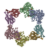
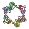
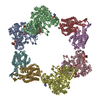
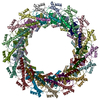
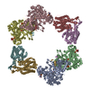
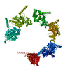
- PDB-5jrj: Crystal Structure of Herbaspirillum seropedicae RecA -

+
Open data


ID or keywords:

Loading...

-
Basic information

Entry
Database: PDB / ID: 5jrj
TitleCrystal Structure of Herbaspirillum seropedicae RecA
ComponentsProtein RecA
KeywordsDNA BINDING PROTEIN / DNA-binding protein / ATP-dependent DNA protein / ATPase activity / DNA strand exchange
Function / homology
Function and homology information


SOS response / ATP-dependent activity, acting on DNA / single-stranded DNA binding / DNA recombination / damaged DNA binding / DNA repair / ATP binding / metal ion binding / cytoplasm
Similarity search - Function
RecA protein, C-terminal domain / Rec A Protein; domain 2 / DNA recombination/repair protein RecA, conserved site / DNA recombination and repair protein RecA, C-terminal / recA signature. / DNA recombination and repair protein RecA / recA bacterial DNA recombination protein / DNA recombination and repair protein RecA, monomer-monomer interface / RecA family profile 2. / DNA recombination and repair protein RecA-like, ATP-binding domain ...RecA protein, C-terminal domain / Rec A Protein; domain 2 / DNA recombination/repair protein RecA, conserved site / DNA recombination and repair protein RecA, C-terminal / recA signature. / DNA recombination and repair protein RecA / recA bacterial DNA recombination protein / DNA recombination and repair protein RecA, monomer-monomer interface / RecA family profile 2. / DNA recombination and repair protein RecA-like, ATP-binding domain / RecA family profile 1. / P-loop containing nucleotide triphosphate hydrolases / ATPases associated with a variety of cellular activities / AAA+ ATPase domain / P-loop containing nucleoside triphosphate hydrolase / Rossmann fold / 2-Layer Sandwich / 3-Layer(aba) Sandwich / Alpha Beta
Similarity search - Domain/homology
ADENOSINE-5'-DIPHOSPHATE / ADENOSINE-5'-TRIPHOSPHATE / Protein RecA / Protein RecA
Similarity search - Component
Biological speciesHerbaspirillum seropedicae (bacteria)
MethodX-RAY DIFFRACTION / MOLECULAR REPLACEMENT / Resolution: 1.7 Å
AuthorsLeite, W.C. / Galvao, C.W. / Saab, S.C. / Iulek, J. / Etto, R.M. / Steffens, M.B.R. / Chitteni-Pattu, S. / Stanage, T. / Keck, J.L. / Cox, M.M.
Funding support United States, 2items
OrganizationGrant numberCountry
National Institutes of Health/National Institute of General Medical Sciences (NIH/NIGMS)GM32335 United States
National Institutes of Health/National Institute of General Medical Sciences (NIH/NIGMS)GM098885 United States
CitationJournal: PLoS ONE / Year: 2016
Title: Structural and Functional Studies of H. seropedicae RecA Protein - Insights into the Polymerization of RecA Protein as Nucleoprotein Filament.
Authors: Leite, W.C. / Galvao, C.W. / Saab, S.C. / Iulek, J. / Etto, R.M. / Steffens, M.B. / Chitteni-Pattu, S. / Stanage, T. / Keck, J.L. / Cox, M.M.
History
DepositionMay 6, 2016Deposition site: RCSB / Processing site: RCSB
Revision 1.0Aug 3, 2016Provider: repository / Type: Initial release
Revision 1.1Sep 13, 2017Group: Author supporting evidence / Derived calculations / Category: pdbx_audit_support / pdbx_struct_oper_list
Item: _pdbx_audit_support.funding_organization / _pdbx_struct_oper_list.symmetry_operation
Revision 1.2Apr 25, 2018Group: Data collection / Database references / Category: citation
Item: _citation.journal_abbrev / _citation.pdbx_database_id_PubMed
Revision 1.3Dec 25, 2019Group: Author supporting evidence / Category: pdbx_audit_support / Item: _pdbx_audit_support.funding_organization
Revision 1.4Sep 27, 2023Group: Data collection / Database references ...Data collection / Database references / Derived calculations / Refinement description
Category: chem_comp_atom / chem_comp_bond ...chem_comp_atom / chem_comp_bond / database_2 / pdbx_initial_refinement_model / pdbx_struct_conn_angle / struct_conn
Item: _database_2.pdbx_DOI / _database_2.pdbx_database_accession ..._database_2.pdbx_DOI / _database_2.pdbx_database_accession / _pdbx_struct_conn_angle.ptnr1_auth_seq_id / _pdbx_struct_conn_angle.ptnr1_symmetry / _pdbx_struct_conn_angle.ptnr3_auth_seq_id / _pdbx_struct_conn_angle.ptnr3_symmetry / _pdbx_struct_conn_angle.value / _struct_conn.pdbx_dist_value / _struct_conn.ptnr2_auth_seq_id / _struct_conn.ptnr2_symmetry

-
Structure visualization

Structure viewerMolecule:
MolmilJmol/JSmol

Downloads & links

-
Assembly

Deposited unit
A: Protein RecA
hetero molecules


Theoretical massNumber of molelcules
Total (without water)38,4914
Polymers37,5171
Non-polymers9743
Water5,855325
1
A: Protein RecA
hetero molecules
x 6


Theoretical massNumber of molelcules
Total (without water)230,94624
Polymers225,0996
Non-polymers5,84718
Water1086
TypeNameSymmetry operationNumber
identity operation1_555x,y,z1
crystal symmetry operation2_555-y,x-y,z+1/31
crystal symmetry operation3_555-x+y,-x,z+2/31
crystal symmetry operation4_555-x,-y,z+1/21
crystal symmetry operation5_555y,-x+y,z+5/61
crystal symmetry operation6_555x-y,x,z+1/61
Unit cell
Length a, b, c (Å)87.410, 87.410, 91.353
Angle α, β, γ (deg.)90.00, 90.00, 120.00
Int Tables number169
Space group name H-MP61
SymmetryHelical symmetry: (Dyad axis: no)

-
Components

#1: Protein Protein RecA / Recombinase A


Mass: 37516.516 Da / Num. of mol.: 1
Source method: isolated from a genetically manipulated source
Source: (gene. exp.) Herbaspirillum seropedicae (bacteria) / Strain: SmR1 / Cell: Proteobacteria / Gene: recA / Production host: Escherichia coli (E. coli) / Strain (production host): STL2669 / References: UniProt: Q9F672, UniProt: D8IYL4*PLUS
#2: Chemical ChemComp-ADP / ADENOSINE-5'-DIPHOSPHATE / Adenosine diphosphate


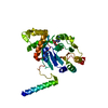
Mass: 427.201 Da / Num. of mol.: 1
Source method: isolated from a genetically manipulated source
Formula: C10H15N5O10P2 / Comment: ADP, energy-carrying molecule*YM
#3: Chemical ChemComp-ATP / ADENOSINE-5'-TRIPHOSPHATE / Adenosine triphosphate


Mass: 507.181 Da / Num. of mol.: 1
Source method: isolated from a genetically manipulated source
Formula: C10H16N5O13P3 / Comment: ATP, energy-carrying molecule*YM
#4: Chemical ChemComp-CA / CALCIUM ION


Mass: 40.078 Da / Num. of mol.: 1
Source method: isolated from a genetically manipulated source
Formula: Ca
#5: Water ChemComp-HOH / water / Water


Mass: 18.015 Da / Num. of mol.: 325 / Source method: isolated from a natural source / Formula: H2O

-
Experimental details

-
Experiment

ExperimentMethod: X-RAY DIFFRACTION / Number of used crystals: 1

-
Sample preparation

CrystalDensity Matthews: 2.69 Å3/Da / Density % sol: 54.2 % / Description: rodlike
Crystal growTemperature: 293.15 K / Method: vapor diffusion, hanging drop / pH: 6.8
Details: Single crystals were obtained with 0.25 M CaCl2 and 14% w/v PEG 3350 with addition of Polypropylene glycol P 400 (PPG 400) onto the drop to give a final concentration of 5-8% w/v.

-
Data collection

DiffractionMean temperature: 100 K
Diffraction sourceSource: ROTATING ANODE / Type: BRUKER AXS MICROSTAR / Wavelength: 1.54 Å
DetectorType: BRUKER SMART 6000 / Detector: CCD / Date: Oct 15, 2014
RadiationProtocol: SINGLE WAVELENGTH / Monochromatic (M) / Laue (L): M / Scattering type: x-ray
Radiation wavelengthWavelength: 1.54 Å / Relative weight: 1
ReflectionResolution: 1.7→31.578 Å / Num. obs: 42574 / % possible obs: 97.67 % / Redundancy: 5.18 % / Biso Wilson estimate: 16.94 Å2 / Rmerge(I) obs: 0.0467 / Net I/σ(I): 31.03
Reflection shellResolution: 1.7→1.73 Å / Redundancy: 1.83 % / Rmerge(I) obs: 0.6285 / Mean I/σ(I) obs: 2.08 / % possible all: 79.5

-
Processing

Software
NameVersionClassification
PHENIX1.9_1692refinement
PROTEUM PLUSVersion 2010.5data reduction
PROTEUM PLUSVersion 2010.5data scaling
PHASERPhaser-2.6.0phasing
Coot0.8.2model building
RefinementMethod to determine structure: MOLECULAR REPLACEMENT
Starting model: 1XMV
Resolution: 1.7→31.578 Å / SU ML: 0.21 / Cross valid method: FREE R-VALUE / Phase error: 22.78
RfactorNum. reflection% reflection
Rfree0.2107 2024 4.75 %
Rwork0.1755 --
obs0.1772 42571 97.66 %
Solvent computationShrinkage radii: 1.3 Å / VDW probe radii: 1.4 Å
Displacement parametersBiso mean: 23.3 Å2
Refinement stepCycle: LAST / Resolution: 1.7→31.578 Å
ProteinNucleic acidLigandSolventTotal
Num. atoms2319 0 0 325 2644
Refine LS restraints
Refine-IDTypeDev idealNumber
X-RAY DIFFRACTIONf_bond_d0.0152453
X-RAY DIFFRACTIONf_angle_d1.6213326
X-RAY DIFFRACTIONf_dihedral_angle_d15.553947
X-RAY DIFFRACTIONf_chiral_restr0.089384
X-RAY DIFFRACTIONf_plane_restr0.007423
LS refinement shell
Resolution (Å)Rfactor RfreeNum. reflection RfreeRfactor RworkNum. reflection RworkRefine-ID% reflection obs (%)
1.6996-1.74210.421090.33482252X-RAY DIFFRACTION76
1.7421-1.78920.30231400.2672732X-RAY DIFFRACTION92
1.7892-1.84180.26411490.22492959X-RAY DIFFRACTION100
1.8418-1.90130.23371480.19892954X-RAY DIFFRACTION100
1.9013-1.96920.22511500.18542935X-RAY DIFFRACTION100
1.9692-2.0480.21551500.18372958X-RAY DIFFRACTION100
2.048-2.14120.22191440.18232963X-RAY DIFFRACTION100
2.1412-2.25410.22361430.17112945X-RAY DIFFRACTION100
2.2541-2.39530.22671470.1692962X-RAY DIFFRACTION100
2.3953-2.58010.20311500.17962966X-RAY DIFFRACTION100
2.5801-2.83960.1991490.17262949X-RAY DIFFRACTION100
2.8396-3.25020.18751470.16422985X-RAY DIFFRACTION100
3.2502-4.09350.18981480.15152971X-RAY DIFFRACTION100
4.0935-31.58350.18551500.15843016X-RAY DIFFRACTION100
Refinement TLS params.Method: refined / Origin x: 33.1732 Å / Origin y: 42.221 Å / Origin z: 42.1896 Å
111213212223313233
T0.1007 Å20.0138 Å2-0.0012 Å2-0.1118 Å20.0134 Å2--0.1049 Å2
L0.593 °2-0.0706 °20.2769 °2-0.2343 °2-0.1199 °2--0.1438 °2
S-0.0415 Å °0.0115 Å °0.0712 Å °-0.0202 Å °-0.0013 Å °-0.0108 Å °-0.0467 Å °0.0006 Å °-0.0319 Å °
Refinement TLS group
IDRefine-IDRefine TLS-IDSelection detailsAuth asym-IDAuth seq-ID
1X-RAY DIFFRACTION1chain A or ( chain A and resid 401 )A6 - 825
2X-RAY DIFFRACTION1chain A or ( chain A and resid 401 )A401

+
About Yorodumi

-
News

-
Feb 9, 2022. New format data for meta-information of EMDB entries

New format data for meta-information of EMDB entries

  • Version 3 of the EMDB header file is now the official format.
  • The previous official version 1.9 will be removed from the archive.

Related info.:EMDB header

External links:wwPDB to switch to version 3 of the EMDB data model

-
Aug 12, 2020. Covid-19 info

Covid-19 info

URL: https://pdbj.org/emnavi/covid19.php

New page: Covid-19 featured information page in EM Navigator.

Related info.:Covid-19 info / Mar 5, 2020. Novel coronavirus structure data

+
Mar 5, 2020. Novel coronavirus structure data

Novel coronavirus structure data

Related info.:Yorodumi Speices / Aug 12, 2020. Covid-19 info

External links:COVID-19 featured content - PDBj / Molecule of the Month (242):Coronavirus Proteases

+
Jan 31, 2019. EMDB accession codes are about to change! (news from PDBe EMDB page)

EMDB accession codes are about to change! (news from PDBe EMDB page)

  • The allocation of 4 digits for EMDB accession codes will soon come to an end. Whilst these codes will remain in use, new EMDB accession codes will include an additional digit and will expand incrementally as the available range of codes is exhausted. The current 4-digit format prefixed with “EMD-” (i.e. EMD-XXXX) will advance to a 5-digit format (i.e. EMD-XXXXX), and so on. It is currently estimated that the 4-digit codes will be depleted around Spring 2019, at which point the 5-digit format will come into force.
  • The EM Navigator/Yorodumi systems omit the EMD- prefix.

Related info.:Q: What is EMD? / ID/Accession-code notation in Yorodumi/EM Navigator

External links:EMDB Accession Codes are Changing Soon! / Contact to PDBj

+
Jul 12, 2017. Major update of PDB

Major update of PDB

  • wwPDB released updated PDB data conforming to the new PDBx/mmCIF dictionary.
  • This is a major update changing the version number from 4 to 5, and with Remediation, in which all the entries are updated.
  • In this update, many items about electron microscopy experimental information are reorganized (e.g. em_software).
  • Now, EM Navigator and Yorodumi are based on the updated data.

External links:wwPDB Remediation / Enriched Model Files Conforming to OneDep Data Standards Now Available in the PDB FTP Archive

-
Yorodumi

Thousand views of thousand structures

  • Yorodumi is a browser for structure data from EMDB, PDB, SASBDB, etc.
  • This page is also the successor to EM Navigator detail page, and also detail information page/front-end page for Omokage search.
  • The word "yorodu" (or yorozu) is an old Japanese word meaning "ten thousand". "mi" (miru) is to see.

Related info.:EMDB / PDB / SASBDB / Comparison of 3 databanks / Yorodumi Search / Aug 31, 2016. New EM Navigator & Yorodumi / Yorodumi Papers / Jmol/JSmol / Function and homology information / Changes in new EM Navigator and Yorodumi

Read more