 Movie
Movie Controller

[English] 日本語
 Yorodumi
- PDB-5jk0: Crystal structure of XerH site-specific recombinase bound to difH... -
+ Open data

- Basic information
| Entry | Database: PDB / ID: 5jk0 | ||||||
|---|---|---|---|---|---|---|---|
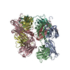
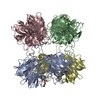
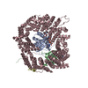
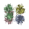
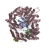
| Title | Crystal structure of XerH site-specific recombinase bound to difH substrate: pre-cleavage complex | ||||||
 Components Components |
| ||||||
 Keywords Keywords |  CELL CYCLE / Xer / tyrosine recombinase / site-specific recombinase / chromosome dimer resolution CELL CYCLE / Xer / tyrosine recombinase / site-specific recombinase / chromosome dimer resolution | ||||||
| Function / homology |  Function and homology information Function and homology informationtyrosine-based site-specific recombinase activity / chromosome segregation / DNA recombination / cell division / DNA binding / cytoplasmSimilarity search - Function | ||||||
| Biological species |   Helicobacter pylori (bacteria)Helicobacter pylori 26695 (bacteria) Helicobacter pylori (bacteria)Helicobacter pylori 26695 (bacteria) | ||||||
| Method | X-RAY DIFFRACTION / SYNCHROTRON / MOLECULAR REPLACEMENT / Resolution: 2.1 Å | ||||||
 Authors Authors | Bebel, A. / Barabas, O. | ||||||
 Citation Citation | Journal: Elife / Year: 2016 Title: Structural snapshots of Xer recombination reveal activation by synaptic complex remodeling and DNA bending. Authors: Bebel, A. / Karaca, E. / Kumar, B. / Stark, W.M. / Barabas, O. | ||||||
| History |
|
- Structure visualization
| Structure viewer | Molecule: MolmilJmol/JSmol |
|---|
- Downloads & links
-Download
| PDBx/mmCIF format | 5jk0.cif.gz | 707.5 KB | Display | PDBx/mmCIF format |
|---|---|---|---|---|
| PDB format | pdb5jk0.ent.gz | 575 KB | Display | PDB format |
| PDBx/mmJSON format | 5jk0.json.gz | Tree view | PDBx/mmJSON format | |
| Others |  Other downloads Other downloads |
-Validation report
| Arichive directory | https://data.pdbj.org/pub/pdb/validation_reports/jk/5jk0ftp://data.pdbj.org/pub/pdb/validation_reports/jk/5jk0 | HTTPS FTP |
|---|
-Related structure data
| Related structure data | 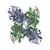 5jjvSC S: Starting model for refinement C: citing same article ( |
|---|---|
| Similar structure data |


-Links
 PDBj
PDBj


- Assembly
| Deposited unit | 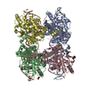
| ||||||||
|---|---|---|---|---|---|---|---|---|---|
| 1 |
| ||||||||
| Unit cell |
|
-Components
-Protein , 1 types, 4 molecules ABCD
| #1: Protein | Mass: 42000.527 Da / Num. of mol.: 4 Source method: isolated from a genetically manipulated source Source: (gene. exp.) Helicobacter pylori (strain ATCC 700392 / 26695) (bacteria)Strain: ATCC 700392 / 26695 / Gene: xerH, HP_0675 / Plasmid: pETM-28 / Production host: Escherichia coli BL21(DE3) (bacteria) / References: UniProt: O25386 |
|---|
-DNA chain , 2 types, 4 molecules EGFH
| #2: DNA chain | Mass: 9195.964 Da / Num. of mol.: 2 / Source method: obtained synthetically / Source: (synth.) Helicobacter pylori 26695 (bacteria)#3: DNA chain | Mass: 9269.026 Da / Num. of mol.: 2 / Source method: obtained synthetically / Source: (synth.) Helicobacter pylori 26695 (bacteria) |
|---|
-Non-polymers , 5 types, 1148 molecules 


| #4: Chemical | ChemComp-GOL / Glycerol Mass: 92.094 Da / Num. of mol.: 5 / Source method: obtained synthetically / Formula: C3H8O3 Mass: 92.094 Da / Num. of mol.: 5 / Source method: obtained synthetically / Formula: C3H8O3#5: Chemical | ChemComp-EDO / Ethylene glycol Mass: 62.068 Da / Num. of mol.: 5 / Source method: obtained synthetically / Formula: C2H6O2 Mass: 62.068 Da / Num. of mol.: 5 / Source method: obtained synthetically / Formula: C2H6O2#6: Chemical | ChemComp-CL / | Chloride Mass: 35.453 Da / Num. of mol.: 1 / Source method: obtained synthetically / Formula: Cl Mass: 35.453 Da / Num. of mol.: 1 / Source method: obtained synthetically / Formula: Cl#7: Chemical | ChemComp-PEG / | Diethylene glycol Mass: 106.120 Da / Num. of mol.: 1 / Source method: obtained synthetically / Formula: C4H10O3 Mass: 106.120 Da / Num. of mol.: 1 / Source method: obtained synthetically / Formula: C4H10O3#8: Water | ChemComp-HOH / | WaterMass: 18.015 Da / Num. of mol.: 1136 / Source method: isolated from a natural source / Formula: H2O |
|---|
-Experimental details
-Experiment
| Experiment | Method: X-RAY DIFFRACTION / Number of used crystals: 1 |
|---|
- Sample preparation
| Crystal | Density Matthews: 2.6 Å3/Da / Density % sol: 52.61 % |
|---|---|
| Crystal grow | Temperature: 279.15 K / Method: vapor diffusion / Details: 0.2M sodium chloride, 21% PEG 3350 |
-Data collection
| Diffraction | Mean temperature: 100 K |
|---|---|
| Diffraction source | Source: SYNCHROTRON / Site: Diamond  / Beamline: I04-1 / Wavelength: 0.92819 Å / Beamline: I04-1 / Wavelength: 0.92819 Å |
| Detector | Type: DECTRIS PILATUS 2M / Detector: PIXEL / Date: Dec 3, 2015 |
| Radiation | Protocol: SINGLE WAVELENGTH / Monochromatic (M) / Laue (L): M / Scattering type: x-ray |
| Radiation wavelength | Wavelength: 0.92819 Å / Relative weight: 1 |
| Reflection | Resolution: 2.1→48.89 Å / Num. obs: 120659 / % possible obs: 99.87 % / Redundancy: 6.6 % / CC1/2: 0.998 / Rsym value: 0.104 / Net I/σ(I): 12.53 |
| Reflection shell | Resolution: 2.1→2.18 Å / Redundancy: 6 % / Mean I/σ(I) obs: 2.25 / % possible all: 99.81 |
- Processing
| Software |
| |||||||||||||||||||||||||||||||||||||||||||||||||||||||||||||||||||||||||||||||||||||||||||||||||||||||||||||||||||||||||||||||||||||||||||||||||||||||||||||||||||||||||||||||||||||||||||||||||||||||||||||||||||||||||
|---|---|---|---|---|---|---|---|---|---|---|---|---|---|---|---|---|---|---|---|---|---|---|---|---|---|---|---|---|---|---|---|---|---|---|---|---|---|---|---|---|---|---|---|---|---|---|---|---|---|---|---|---|---|---|---|---|---|---|---|---|---|---|---|---|---|---|---|---|---|---|---|---|---|---|---|---|---|---|---|---|---|---|---|---|---|---|---|---|---|---|---|---|---|---|---|---|---|---|---|---|---|---|---|---|---|---|---|---|---|---|---|---|---|---|---|---|---|---|---|---|---|---|---|---|---|---|---|---|---|---|---|---|---|---|---|---|---|---|---|---|---|---|---|---|---|---|---|---|---|---|---|---|---|---|---|---|---|---|---|---|---|---|---|---|---|---|---|---|---|---|---|---|---|---|---|---|---|---|---|---|---|---|---|---|---|---|---|---|---|---|---|---|---|---|---|---|---|---|---|---|---|---|---|---|---|---|---|---|---|---|---|---|---|---|---|---|---|---|
| Refinement | Method to determine structure: MOLECULAR REPLACEMENT Starting model: 5JJV Resolution: 2.1→48.89 Å / SU ML: 0.26 / Cross valid method: FREE R-VALUE / σ(F): 1.35 / Phase error: 23.26
| |||||||||||||||||||||||||||||||||||||||||||||||||||||||||||||||||||||||||||||||||||||||||||||||||||||||||||||||||||||||||||||||||||||||||||||||||||||||||||||||||||||||||||||||||||||||||||||||||||||||||||||||||||||||||
| Solvent computation | Shrinkage radii: 0.9 Å / VDW probe radii: 1.11 Å | |||||||||||||||||||||||||||||||||||||||||||||||||||||||||||||||||||||||||||||||||||||||||||||||||||||||||||||||||||||||||||||||||||||||||||||||||||||||||||||||||||||||||||||||||||||||||||||||||||||||||||||||||||||||||
| Refinement step | Cycle: LAST / Resolution: 2.1→48.89 Å
| |||||||||||||||||||||||||||||||||||||||||||||||||||||||||||||||||||||||||||||||||||||||||||||||||||||||||||||||||||||||||||||||||||||||||||||||||||||||||||||||||||||||||||||||||||||||||||||||||||||||||||||||||||||||||
| Refine LS restraints |
| |||||||||||||||||||||||||||||||||||||||||||||||||||||||||||||||||||||||||||||||||||||||||||||||||||||||||||||||||||||||||||||||||||||||||||||||||||||||||||||||||||||||||||||||||||||||||||||||||||||||||||||||||||||||||
| LS refinement shell |
|
