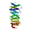
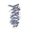
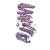
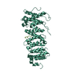
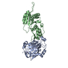
mitotic checkpoint complex / protein phosphatase type 2A complex / meiotic sister chromatid cohesion, centromeric / Inactivation of APC/C via direct inhibition of the APC/C complex / APC/C:Cdc20 mediated degradation of mitotic proteins / anaphase-promoting complex / metaphase/anaphase transition of mitotic cell cycle / protein phosphatase regulator activity / outer kinetochore / protein localization to chromosome, centromeric region ...mitotic checkpoint complex / protein phosphatase type 2A complex / meiotic sister chromatid cohesion, centromeric / Inactivation of APC/C via direct inhibition of the APC/C complex / APC/C:Cdc20 mediated degradation of mitotic proteins / anaphase-promoting complex / metaphase/anaphase transition of mitotic cell cycle / protein phosphatase regulator activity / outer kinetochore / protein localization to chromosome, centromeric region / APC truncation mutants have impaired AXIN binding / AXIN missense mutants destabilize the destruction complex / Truncations of AMER1 destabilize the destruction complex / Beta-catenin phosphorylation cascade / Signaling by GSK3beta mutants / CTNNB1 S33 mutants aren't phosphorylated / CTNNB1 S37 mutants aren't phosphorylated / CTNNB1 S45 mutants aren't phosphorylated / CTNNB1 T41 mutants aren't phosphorylated / Disassembly of the destruction complex and recruitment of AXIN to the membrane / CTLA4 inhibitory signaling / Platelet sensitization by LDL / mitotic spindle assembly checkpoint signaling / protein phosphatase activator activity / chromosome, centromeric region / intrinsic apoptotic signaling pathway in response to DNA damage by p53 class mediator / Amplification of signal from unattached kinetochores via a MAD2 inhibitory signal / DNA damage response, signal transduction by p53 class mediator resulting in cell cycle arrest / Mitotic Prometaphase / EML4 and NUDC in mitotic spindle formation / Resolution of Sister Chromatid Cohesion / APC-Cdc20 mediated degradation of Nek2A / RHO GTPases Activate Formins / Cdc20:Phospho-APC/C mediated degradation of Cyclin A / RAF activation / Degradation of beta-catenin by the destruction complex / kinetochore / spindle / Negative regulation of MAPK pathway / Separation of Sister Chromatids / Regulation of TP53 Degradation / PI5P, PP2A and IER3 Regulate PI3K/AKT Signaling / proteasome-mediated ubiquitin-dependent protein catabolic process / non-specific serine/threonine protein kinase / protein kinase activity / cell division / negative regulation of cell population proliferation / phosphorylation / protein serine kinase activity / protein serine/threonine kinase activity / centrosome / apoptotic process / perinuclear region of cytoplasm / Golgi apparatus / signal transduction / nucleoplasm / ATP binding / nucleus / cytosol / cytoplasm Similarity search - Function
Mad3/Bub1 homology region 1 / Mitotic spindle checkpoint protein Bub1/Mad3 / Mad3/BUB1 homology region 1 / BUB1 N-terminal domain profile. / Mad3/BUB1 hoMad3/BUB1 homology region 1 / Protein phosphatase 2A, regulatory B subunit, B56 / Protein phosphatase 2A regulatory B subunit (B56 family) / Leucine-rich Repeat Variant / Leucine-rich Repeat Variant / Armadillo-like helical ...Mad3/Bub1 homology region 1 / Mitotic spindle checkpoint protein Bub1/Mad3 / Mad3/BUB1 homology region 1 / BUB1 N-terminal domain profile. / Mad3/BUB1 hoMad3/BUB1 homology region 1 / Protein phosphatase 2A, regulatory B subunit, B56 / Protein phosphatase 2A regulatory B subunit (B56 family) / Leucine-rich Repeat Variant / Leucine-rich Repeat Variant / Armadillo-like helical / Alpha Horseshoe / Armadillo-type fold / Protein kinase-like domain superfamily / Mainly Alpha Similarity search - Domain/homology
Resolution: 2.35→50 Å / Cor.coef. Fo:Fc: 0.954 / Cor.coef. Fo:Fc free: 0.943 / SU B: 8.913 / SU ML: 0.108 / Cross valid method: THROUGHOUT / σ(F): 0 / ESU R: 0.188 / ESU R Free: 0.161 Details: HYDROGENS HAVE BEEN ADDED IN THE RIDING POSITIONS U VALUES : WITH TLS ADDED
Rfactor
Num. reflection
% reflection
Selection details
Rfree
0.2041
2882
5 %
RANDOM
Rwork
0.1795
-
-
-
obs
0.1807
54953
99.53 %
-
Solvent computation
Ion probe radii: 0.8 Å / Shrinkage radii: 0.8 Å / VDW probe radii: 1.2 Å
In the structure databanks used in Yorodumi, some data are registered as the other names, "COVID-19 virus" and "2019-nCoV". Here are the details of the virus and the list of structure data.
Jan 31, 2019. EMDB accession codes are about to change! (news from PDBe EMDB page)
EMDB accession codes are about to change! (news from PDBe EMDB page)
The allocation of 4 digits for EMDB accession codes will soon come to an end. Whilst these codes will remain in use, new EMDB accession codes will include an additional digit and will expand incrementally as the available range of codes is exhausted. The current 4-digit format prefixed with “EMD-” (i.e. EMD-XXXX) will advance to a 5-digit format (i.e. EMD-XXXXX), and so on. It is currently estimated that the 4-digit codes will be depleted around Spring 2019, at which point the 5-digit format will come into force.
The EM Navigator/Yorodumi systems omit the EMD- prefix.
Related info.:Q: What is EMD? / ID/Accession-code notation in Yorodumi/EM Navigator
Yorodumi is a browser for structure data from EMDB, PDB, SASBDB, etc.
This page is also the successor to EM Navigator detail page, and also detail information page/front-end page for Omokage search.
The word "yorodu" (or yorozu) is an old Japanese word meaning "ten thousand". "mi" (miru) is to see.
Related info.:EMDB / PDB / SASBDB / Comparison of 3 databanks / Yorodumi Search / Aug 31, 2016. New EM Navigator & Yorodumi / Yorodumi Papers / Jmol/JSmol / Function and homology information / Changes in new EM Navigator and Yorodumi
 Movie
Movie Controller


 Open data
 Basic information
 Components
 Keywords
 SIGNALING PROTEIN /
  Function and homology information
 Authors
 China, 2items
  Citation
 Structure visualization
 Downloads & links
 Other downloads


 PDBj
PDBj


 Assembly
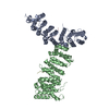

 Mass: 18.015 Da / Num. of mol.: 240 / Source method: isolated from a natural source / Formula: H2O
 Sample preparation
 Processing