 Movie
Movie Controller

[English] 日本語
 Yorodumi
- PDB-5jdw: CRYSTAL STRUCTURE OF HUMAN L-ARGININE:GLYCINE AMIDINOTRANSFERASE ... -
+ Open data

- Basic information
| Entry | Database: PDB / ID: 5jdw | ||||||
|---|---|---|---|---|---|---|---|
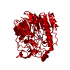
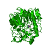
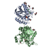
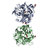
| Title | CRYSTAL STRUCTURE OF HUMAN L-ARGININE:GLYCINE AMIDINOTRANSFERASE IN COMPLEX WITH GLYCINE | ||||||
 Components Components | PROTEIN (L-ARGININE:GLYCINE AMIDINOTRANSFERASE) | ||||||
 Keywords Keywords |  TRANSFERASE / CREATINE BIOSYNTHESIS / CATALYTIC TRIAD / REACTION MECHANISM / NOVEL FOLD / FIVEFOLD PSEUDOSYMMETRY TRANSFERASE / CREATINE BIOSYNTHESIS / CATALYTIC TRIAD / REACTION MECHANISM / NOVEL FOLD / FIVEFOLD PSEUDOSYMMETRY | ||||||
| Function / homology |  Function and homology informationglycine amidinotransferase / glycine amidinotransferase activity / amidinotransferase activity / creatine metabolic process / Creatine metabolism / creatine biosynthetic process / muscle atrophy / mitochondrial intermembrane space / positive regulation of cold-induced thermogenesis / mitochondrial inner membrane ...glycine amidinotransferase / glycine amidinotransferase activity / amidinotransferase activity / creatine metabolic process / Creatine metabolism / creatine biosynthetic process / muscle atrophy / mitochondrial intermembrane space / positive regulation of cold-induced thermogenesis / mitochondrial inner membrane / learning or memory / mitochondrion / extracellular exosome Function and homology informationglycine amidinotransferase / glycine amidinotransferase activity / amidinotransferase activity / creatine metabolic process / Creatine metabolism / creatine biosynthetic process / muscle atrophy / mitochondrial intermembrane space / positive regulation of cold-induced thermogenesis / mitochondrial inner membrane ...glycine amidinotransferase / glycine amidinotransferase activity / amidinotransferase activity / creatine metabolic process / Creatine metabolism / creatine biosynthetic process / muscle atrophy / mitochondrial intermembrane space / positive regulation of cold-induced thermogenesis / mitochondrial inner membrane / learning or memory / mitochondrion / extracellular exosomeSimilarity search - Function | ||||||
| Biological species |  Homo sapiens (human) Homo sapiens (human) | ||||||
| Method | X-RAY DIFFRACTION / OTHER / Resolution: 2.6 Å | ||||||
 Authors Authors | Fritsche, E. / Humm, A. / Huber, R. | ||||||
 Citation Citation | Journal: J.Biol.Chem. / Year: 1999 Title: The ligand-induced structural changes of human L-Arginine:Glycine amidinotransferase. A mutational and crystallographic study. Authors: Fritsche, E. / Humm, A. / Huber, R. | ||||||
| History |
|
- Structure visualization
| Structure viewer | Molecule: MolmilJmol/JSmol |
|---|
- Downloads & links
-Download
| PDBx/mmCIF format | 5jdw.cif.gz | 106.4 KB | Display | PDBx/mmCIF format |
|---|---|---|---|---|
| PDB format | pdb5jdw.ent.gz | 82.9 KB | Display | PDB format |
| PDBx/mmJSON format | 5jdw.json.gz | Tree view | PDBx/mmJSON format | |
| Others |  Other downloads Other downloads |
-Validation report
| Arichive directory | https://data.pdbj.org/pub/pdb/validation_reports/jd/5jdwftp://data.pdbj.org/pub/pdb/validation_reports/jd/5jdw | HTTPS FTP |
|---|
-Related structure data
| Related structure data |  1jdxC 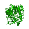 2jdxC  6jdwC  7jdwC  8jdwC 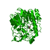 9jdwC C: citing same article ( |
|---|---|
| Similar structure data |


-Links
 PDBj
PDBj

- Assembly
| Deposited unit | 
| ||||||||
|---|---|---|---|---|---|---|---|---|---|
| 1 | 
| ||||||||
| Unit cell |
|
-Components
| #1: Protein | Mass: 44341.488 Da / Num. of mol.: 1 / Fragment: RESIDUES 38 - 423 Source method: isolated from a genetically manipulated source Source: (gene. exp.) Homo sapiens (human) / Strain: BL(21)DE3PLYSS / Cellular location: CYTOSOLICCytosol / Gene: AT38H / Organ: KIDNEY / Organelle: MITOCHONDRIAMitochondrion / Plasmid: PRSETAT38H / Gene (production host): AT38H / Production host:  Escherichia coli (E. coli) / Strain (production host): BL21 (DE3) PLYSS / References: UniProt: P50440, glycine amidinotransferase Escherichia coli (E. coli) / Strain (production host): BL21 (DE3) PLYSS / References: UniProt: P50440, glycine amidinotransferase |
|---|---|
| #2: Chemical | ChemComp-GLY / Glycine  Type: peptide linking / Mass: 75.067 Da / Num. of mol.: 1 / Source method: obtained synthetically / Formula: C2H5NO2 Type: peptide linking / Mass: 75.067 Da / Num. of mol.: 1 / Source method: obtained synthetically / Formula: C2H5NO2 |
| #3: Water | ChemComp-HOH / Water Mass: 18.015 Da / Num. of mol.: 145 / Source method: isolated from a natural source / Formula: H2O Mass: 18.015 Da / Num. of mol.: 145 / Source method: isolated from a natural source / Formula: H2O |
-Experimental details
-Experiment
| Experiment | Method: X-RAY DIFFRACTION / Number of used crystals: 1 |
|---|
- Sample preparation
| Crystal | Density Matthews: 3.83 Å3/Da / Density % sol: 68 % | |||||||||||||||||||||||||||||||||||
|---|---|---|---|---|---|---|---|---|---|---|---|---|---|---|---|---|---|---|---|---|---|---|---|---|---|---|---|---|---|---|---|---|---|---|---|---|
| Crystal grow | pH: 7 / Details: pH 7.0 | |||||||||||||||||||||||||||||||||||
| Crystal grow | *PLUS Method: vapor diffusion, hanging dropDetails: drop was made of a 7 micro litter protein solution and 14 micro litter of a reservoir solution | |||||||||||||||||||||||||||||||||||
| Components of the solutions | *PLUS
|
-Data collection
| Diffraction | Mean temperature: 293 K |
|---|---|
| Diffraction source | Source: ROTATING ANODE / Type: RIGAKU RU200 / Wavelength: 1.5418 |
| Detector | Type: MARRESEARCH / Detector: IMAGE PLATE |
| Radiation | Protocol: SINGLE WAVELENGTH / Monochromatic (M) / Laue (L): M / Scattering type: x-ray |
| Radiation wavelength | Wavelength: 1.5418 Å / Relative weight: 1 |
| Reflection | Resolution: 2.6→20 Å / Num. obs: 20930 / % possible obs: 93.7 % / Observed criterion σ(I): 2 / Redundancy: 3.4 % / Rsym value: 0.115 |
- Processing
| Software |
| ||||||||||||||||||||||||||||||||||||||||||||||||||||||||||||
|---|---|---|---|---|---|---|---|---|---|---|---|---|---|---|---|---|---|---|---|---|---|---|---|---|---|---|---|---|---|---|---|---|---|---|---|---|---|---|---|---|---|---|---|---|---|---|---|---|---|---|---|---|---|---|---|---|---|---|---|---|---|
| Refinement | Method to determine structure: OTHER / Resolution: 2.6→8 Å / Cross valid method: FREE R / σ(F): 0
| ||||||||||||||||||||||||||||||||||||||||||||||||||||||||||||
| Refinement step | Cycle: LAST / Resolution: 2.6→8 Å
| ||||||||||||||||||||||||||||||||||||||||||||||||||||||||||||
| Refine LS restraints |
|
