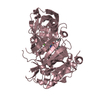
 Movie
Movie Controller


+ Open data

- Basic information
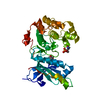
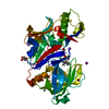
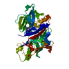
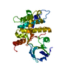
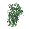
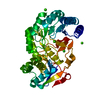
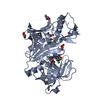
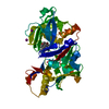
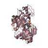
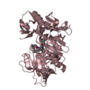
| Entry | Database: PDB / ID: 5j8i | ||||||
|---|---|---|---|---|---|---|---|
| Title | Crystal structure of TL11-113 bound to TAK1-TAB1 | ||||||
 Components Components | Mitogen-activated protein kinase kinase kinase 7/TGF-beta-activated kinase 1 and MAP3K7-binding protein 1 chimera | ||||||
 Keywords Keywords | TRANSFERASE/TRANSFERASE inhibitor / Mitogen-activated protein kinase kinase kinase 7/TGF-beta-activated kinase 1 and MAP3K7-binding protein 1 / TRANSFERASE-TRANSFERASE inhibitor complex | ||||||
| Function / homology |  Function and homology information Function and homology informationpositive regulation of cGAS/STING signaling pathway / histone kinase activity /  MAP kinase kinase kinase kinase activity / nucleotide-binding domain, leucine rich repeat containing receptor signaling pathway / linear polyubiquitin binding / interleukin-17A-mediated signaling pathway / cardiac septum development / I-kappaB phosphorylation / mitogen-activated protein kinase kinase kinase / interleukin-33-mediated signaling pathway ...positive regulation of cGAS/STING signaling pathway / histone kinase activity / MAP kinase kinase kinase kinase activity / nucleotide-binding domain, leucine rich repeat containing receptor signaling pathway / linear polyubiquitin binding / interleukin-17A-mediated signaling pathway / cardiac septum development / I-kappaB phosphorylation / mitogen-activated protein kinase kinase kinase / interleukin-33-mediated signaling pathway / toll-like receptor 3 signaling pathway / type II transforming growth factor beta receptor binding / TRIF-dependent toll-like receptor signaling pathway / activation of NF-kappaB-inducing kinase activity / coronary vasculature development / ATAC complex / positive regulation of vascular associated smooth muscle cell migration / cellular response to angiotensin / anoikis / aorta development / MyD88-dependent toll-like receptor signaling pathway / interleukin-1-mediated signaling pathway / toll-like receptor 4 signaling pathway / protein serine/threonine kinase binding / protein serine/threonine phosphatase activity / mitogen-activated protein kinase p38 binding / cytoplasmic pattern recognition receptor signaling pathway / p38MAPK cascade / non-canonical NF-kappaB signal transduction / Fc-epsilon receptor signaling pathway / stimulatory C-type lectin receptor signaling pathway / positive regulation of macroautophagy / positive regulation of cell size / MAP kinase kinase kinase activity / MAP kinase activity / canonical NF-kappaB signal transduction / enzyme activator activity / positive regulation of cell cycle / stress-activated MAPK cascade / heart morphogenesis / positive regulation of JUN kinase activity / JNK cascade / IRAK2 mediated activation of TAK1 complex / Alpha-protein kinase 1 signaling pathway / positive regulation of vascular associated smooth muscle cell proliferation / IRAK2 mediated activation of TAK1 complex upon TLR7/8 or 9 stimulation / TICAM1,TRAF6-dependent induction of TAK1 complex / positive regulation of interleukin-2 production / TRAF6-mediated induction of TAK1 complex within TLR4 complex / protein serine/threonine kinase activator activity / transforming growth factor beta receptor signaling pathway / JNK (c-Jun kinases) phosphorylation and activation mediated by activated human TAK1 / TNFR1-induced NF-kappa-B signaling pathway / activated TAK1 mediates p38 MAPK activation / Activation of NF-kappaB in B cells / TAK1-dependent IKK and NF-kappa-B activation / lung development / NOD1/2 Signaling Pathway / positive regulation of protein serine/threonine kinase activity / receptor tyrosine kinase binding / positive regulation of T cell cytokine production / CLEC7A (Dectin-1) signaling / transcription coactivator binding / FCERI mediated NF-kB activation / Interleukin-1 signaling / positive regulation of non-canonical NF-kappaB signal transduction / MAPK cascade / Downstream TCR signaling / Ca2+ pathway / cellular response to tumor necrosis factor / T cell receptor signaling pathway / cellular response to hypoxia / scaffold protein binding / DNA-binding transcription factor binding / positive regulation of canonical NF-kappaB signal transduction / in utero embryonic development / positive regulation of MAPK cascade / molecular adaptor activity / endosome membrane / Ub-specific processing proteases / nuclear speck / defense response to bacterium / inflammatory response / immune response / negative regulation of gene expression / protein serine kinase activity / protein serine/threonine kinase activity / ubiquitin protein ligase binding / protein-containing complex binding / endoplasmic reticulum membrane / SARS-CoV-2 activates/modulates innate and adaptive immune responses / magnesium ion binding / endoplasmic reticulum / protein-containing complex / ATP binding / identical protein binding / nucleus / plasma membrane / cytosol / cytoplasm MAP kinase kinase kinase kinase activity / nucleotide-binding domain, leucine rich repeat containing receptor signaling pathway / linear polyubiquitin binding / interleukin-17A-mediated signaling pathway / cardiac septum development / I-kappaB phosphorylation / mitogen-activated protein kinase kinase kinase / interleukin-33-mediated signaling pathway ...positive regulation of cGAS/STING signaling pathway / histone kinase activity / MAP kinase kinase kinase kinase activity / nucleotide-binding domain, leucine rich repeat containing receptor signaling pathway / linear polyubiquitin binding / interleukin-17A-mediated signaling pathway / cardiac septum development / I-kappaB phosphorylation / mitogen-activated protein kinase kinase kinase / interleukin-33-mediated signaling pathway / toll-like receptor 3 signaling pathway / type II transforming growth factor beta receptor binding / TRIF-dependent toll-like receptor signaling pathway / activation of NF-kappaB-inducing kinase activity / coronary vasculature development / ATAC complex / positive regulation of vascular associated smooth muscle cell migration / cellular response to angiotensin / anoikis / aorta development / MyD88-dependent toll-like receptor signaling pathway / interleukin-1-mediated signaling pathway / toll-like receptor 4 signaling pathway / protein serine/threonine kinase binding / protein serine/threonine phosphatase activity / mitogen-activated protein kinase p38 binding / cytoplasmic pattern recognition receptor signaling pathway / p38MAPK cascade / non-canonical NF-kappaB signal transduction / Fc-epsilon receptor signaling pathway / stimulatory C-type lectin receptor signaling pathway / positive regulation of macroautophagy / positive regulation of cell size / MAP kinase kinase kinase activity / MAP kinase activity / canonical NF-kappaB signal transduction / enzyme activator activity / positive regulation of cell cycle / stress-activated MAPK cascade / heart morphogenesis / positive regulation of JUN kinase activity / JNK cascade / IRAK2 mediated activation of TAK1 complex / Alpha-protein kinase 1 signaling pathway / positive regulation of vascular associated smooth muscle cell proliferation / IRAK2 mediated activation of TAK1 complex upon TLR7/8 or 9 stimulation / TICAM1,TRAF6-dependent induction of TAK1 complex / positive regulation of interleukin-2 production / TRAF6-mediated induction of TAK1 complex within TLR4 complex / protein serine/threonine kinase activator activity / transforming growth factor beta receptor signaling pathway / JNK (c-Jun kinases) phosphorylation and activation mediated by activated human TAK1 / TNFR1-induced NF-kappa-B signaling pathway / activated TAK1 mediates p38 MAPK activation / Activation of NF-kappaB in B cells / TAK1-dependent IKK and NF-kappa-B activation / lung development / NOD1/2 Signaling Pathway / positive regulation of protein serine/threonine kinase activity / receptor tyrosine kinase binding / positive regulation of T cell cytokine production / CLEC7A (Dectin-1) signaling / transcription coactivator binding / FCERI mediated NF-kB activation / Interleukin-1 signaling / positive regulation of non-canonical NF-kappaB signal transduction / MAPK cascade / Downstream TCR signaling / Ca2+ pathway / cellular response to tumor necrosis factor / T cell receptor signaling pathway / cellular response to hypoxia / scaffold protein binding / DNA-binding transcription factor binding / positive regulation of canonical NF-kappaB signal transduction / in utero embryonic development / positive regulation of MAPK cascade / molecular adaptor activity / endosome membrane / Ub-specific processing proteases / nuclear speck / defense response to bacterium / inflammatory response / immune response / negative regulation of gene expression / protein serine kinase activity / protein serine/threonine kinase activity / ubiquitin protein ligase binding / protein-containing complex binding / endoplasmic reticulum membrane / SARS-CoV-2 activates/modulates innate and adaptive immune responses / magnesium ion binding / endoplasmic reticulum / protein-containing complex / ATP binding / identical protein binding / nucleus / plasma membrane / cytosol / cytoplasmSimilarity search - Function | ||||||
| Biological species |  Homo sapiens (human) Homo sapiens (human) | ||||||
| Method | X-RAY DIFFRACTION / SYNCHROTRON / MOLECULAR REPLACEMENT / Resolution: 2.404 Å | ||||||
 Authors Authors | Gurbani, D. / Westover, K.D. | ||||||
| Funding support |  United States, 1items United States, 1items
| ||||||
 Citation Citation | Journal: Bioorg. Med. Chem. / Year: 2017 Title: Structure-guided development of covalent TAK1 inhibitors. Authors: Tan, L. / Gurbani, D. / Weisberg, E.L. / Hunter, J.C. / Li, L. / Jones, D.S. / Ficarro, S.B. / Mowafy, S. / Tam, C.P. / Rao, S. / Du, G. / Griffin, J.D. / Sorger, P.K. / Marto, J.A. / Westover, K.D. / Gray, N.S. | ||||||
| History |
|
- Structure visualization
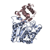
| Structure viewer | Molecule: MolmilJmol/JSmol |
|---|
- Downloads & links
-Download
| PDBx/mmCIF format | 5j8i.cif.gz | 122.4 KB | Display | PDBx/mmCIF format |
|---|---|---|---|---|
| PDB format | pdb5j8i.ent.gz | 95.1 KB | Display | PDB format |
| PDBx/mmJSON format | 5j8i.json.gz | Tree view | PDBx/mmJSON format | |
| Others |  Other downloads Other downloads |
-Validation report
| Arichive directory | https://data.pdbj.org/pub/pdb/validation_reports/j8/5j8iftp://data.pdbj.org/pub/pdb/validation_reports/j8/5j8i | HTTPS FTP |
|---|
-Related structure data
| Related structure data | 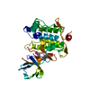 5e7rC 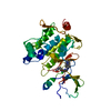 5j7sC 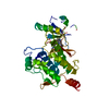 5j9lC 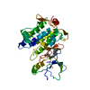 5jh6C 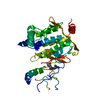 5jk3C  2yiyS C: citing same article ( S: Starting model for refinement |
|---|---|
| Similar structure data |


-Links
 PDBj
PDBj


- Assembly
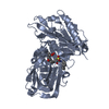
| Deposited unit | 
| ||||||||
|---|---|---|---|---|---|---|---|---|---|
| 1 |
| ||||||||
| Unit cell |
|
-Components
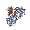
| #1: Protein | Mass: 35463.867 Da / Num. of mol.: 1 Fragment: UNP O43318 residues 31-303, Q15750 residues 468-504 Source method: isolated from a genetically manipulated source Source: (gene. exp.) Homo sapiens (human) / Gene: MAP3K7, TAK1, TAB1, MAP3K7IP1 / Production host:   Spodoptera frugiperda (fall armyworm) Spodoptera frugiperda (fall armyworm)References: UniProt: O43318, UniProt: Q15750, mitogen-activated protein kinase kinase kinase |
|---|---|
| #2: Chemical | ChemComp-6H4 /   Mass: 464.947 Da / Num. of mol.: 1 / Source method: obtained synthetically / Formula: C24H25ClN6O2 Mass: 464.947 Da / Num. of mol.: 1 / Source method: obtained synthetically / Formula: C24H25ClN6O2 |
-Experimental details
-Experiment
| Experiment | Method: X-RAY DIFFRACTION / Number of used crystals: 1 |
|---|
- Sample preparation
| Crystal | Density Matthews: 3.97 Å3/Da / Density % sol: 69.02 % |
|---|---|
| Crystal grow | Temperature: 293 K / Method: vapor diffusion, hanging drop / pH: 7 Details: 0.75M NaCitrate, 0.1M Tris-HCL, 0.2M NaCl, pH 7.0, 5mM Adenosine; |
-Data collection
| Diffraction | Mean temperature: 100 K |
|---|---|
| Diffraction source | Source: SYNCHROTRON / Site: APS / Beamline: 19-ID / Wavelength: 0.97932 Å |
| Detector | Type: ADSC QUANTUM 315 / Detector: CCD / Date: Oct 15, 2015 |
| Radiation | Monochromator: Rosenbaum-Rock high-resolution double-crystal monochromator Protocol: SINGLE WAVELENGTH / Monochromatic (M) / Laue (L): M / Scattering type: x-ray |
| Radiation wavelength | Wavelength: 0.97932 Å / Relative weight: 1 |
| Reflection | Resolution: 2.43→50 Å / Num. obs: 21238 / % possible obs: 96.3 % / Redundancy: 8.3 % / Rmerge(I) obs: 0.124 / Net I/σ(I): 18.94 |
| Reflection shell | Resolution: 2.43→2.47 Å / Redundancy: 5.6 % / Rmerge(I) obs: 1.571 / Mean I/σ(I) obs: 1.16 / % possible all: 80.1 |
- Processing
| Software |
| |||||||||||||||||||||||||||||||||||||||||||||||||||||||||||||||
|---|---|---|---|---|---|---|---|---|---|---|---|---|---|---|---|---|---|---|---|---|---|---|---|---|---|---|---|---|---|---|---|---|---|---|---|---|---|---|---|---|---|---|---|---|---|---|---|---|---|---|---|---|---|---|---|---|---|---|---|---|---|---|---|---|
| Refinement | Method to determine structure: MOLECULAR REPLACEMENT Starting model: 2YIY Resolution: 2.404→49.081 Å / SU ML: 0.36 / Cross valid method: FREE R-VALUE / σ(F): 1.35 / Phase error: 33.58 / Stereochemistry target values: ML
| |||||||||||||||||||||||||||||||||||||||||||||||||||||||||||||||
| Solvent computation | Shrinkage radii: 0.9 Å / VDW probe radii: 1.11 Å / Solvent model: FLAT BULK SOLVENT MODEL | |||||||||||||||||||||||||||||||||||||||||||||||||||||||||||||||
| Refinement step | Cycle: LAST / Resolution: 2.404→49.081 Å
| |||||||||||||||||||||||||||||||||||||||||||||||||||||||||||||||
| Refine LS restraints |
| |||||||||||||||||||||||||||||||||||||||||||||||||||||||||||||||
| LS refinement shell |
|
