 Movie
Movie Controller


+ Open data

- Basic information
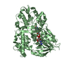
| Entry | Database: PDB / ID: 5j7x | ||||||
|---|---|---|---|---|---|---|---|
| Title | Baeyer-Villiger monooxygenase BVMOAFL838 from Aspergillus flavus | ||||||
 Components Components | Dimethylaniline monooxygenase, putative | ||||||
 Keywords Keywords |  OXIDOREDUCTASE / Baeyer-Villiger monooxygenase OXIDOREDUCTASE / Baeyer-Villiger monooxygenase | ||||||
| Function / homology |  Function and homology information Function and homology informationN,N-dimethylaniline monooxygenase activity / NADP binding / flavin adenine dinucleotide binding / membrane => GO:0016020Similarity search - Function | ||||||
| Biological species |  Aspergillus flavus (mold) Aspergillus flavus (mold) | ||||||
| Method | X-RAY DIFFRACTION / SYNCHROTRON / MOLECULAR REPLACEMENT / molecular replacement / Resolution: 1.9 Å | ||||||
 Authors Authors | Ferroni, F.M. / Tolmie, C. / Smit, M.S. / Opperman, D.J. | ||||||
 Citation Citation | Journal: Plos One / Year: 2016 Title: Structural and Catalytic Characterization of a Fungal Baeyer-Villiger Monooxygenase. Authors: Ferroni, F.M. / Tolmie, C. / Smit, M.S. / Opperman, D.J. | ||||||
| History |
|
- Structure visualization
| Structure viewer | Molecule: MolmilJmol/JSmol |
|---|
- Downloads & links
-Download
| PDBx/mmCIF format | 5j7x.cif.gz | 241.5 KB | Display | PDBx/mmCIF format |
|---|---|---|---|---|
| PDB format | pdb5j7x.ent.gz | 192.5 KB | Display | PDB format |
| PDBx/mmJSON format | 5j7x.json.gz | Tree view | PDBx/mmJSON format | |
| Others |  Other downloads Other downloads |
-Validation report
| Arichive directory | https://data.pdbj.org/pub/pdb/validation_reports/j7/5j7xftp://data.pdbj.org/pub/pdb/validation_reports/j7/5j7x | HTTPS FTP |
|---|
-Related structure data
| Related structure data |  1w4xS S: Starting model for refinement |
|---|---|
| Similar structure data |


-Links
 PDBj
PDBj- Assembly
| Deposited unit | 
| ||||||||
|---|---|---|---|---|---|---|---|---|---|
| 1 |
| ||||||||
| Unit cell |
|
-Components
| #1: Protein | Mass: 62241.844 Da / Num. of mol.: 1 Source method: isolated from a genetically manipulated source Source: (gene. exp.) Aspergillus flavus (mold) / Gene: AFLA_016370 / Plasmid: pET22b(+) / Production host:  Escherichia coli BL21(DE3) (bacteria) / References: UniProt: B8N653 Escherichia coli BL21(DE3) (bacteria) / References: UniProt: B8N653 |
|---|---|
| #2: Chemical | ChemComp-FAD / Flavin adenine dinucleotide  Mass: 785.550 Da / Num. of mol.: 1 / Source method: obtained synthetically / Formula: C27H33N9O15P2 / Comment: FAD*YM Mass: 785.550 Da / Num. of mol.: 1 / Source method: obtained synthetically / Formula: C27H33N9O15P2 / Comment: FAD*YM |
| #3: Chemical | ChemComp-SO4 / Sulfate  Mass: 96.063 Da / Num. of mol.: 1 / Source method: obtained synthetically / Formula: SO4 Mass: 96.063 Da / Num. of mol.: 1 / Source method: obtained synthetically / Formula: SO4 |
| #4: Chemical | ChemComp-TAM / Tris  Mass: 163.215 Da / Num. of mol.: 1 / Source method: obtained synthetically / Formula: C7H17NO3 / Comment: pH buffer*YM Mass: 163.215 Da / Num. of mol.: 1 / Source method: obtained synthetically / Formula: C7H17NO3 / Comment: pH buffer*YM |
| #5: Water | ChemComp-HOH / Water Mass: 18.015 Da / Num. of mol.: 361 / Source method: isolated from a natural source / Formula: H2O Mass: 18.015 Da / Num. of mol.: 361 / Source method: isolated from a natural source / Formula: H2O |
-Experimental details
-Experiment
| Experiment | Method: X-RAY DIFFRACTION / Number of used crystals: 1 |
|---|
- Sample preparation
| Crystal | Density Matthews: 2.99 Å3/Da / Density % sol: 58.81 % |
|---|---|
| Crystal grow | Temperature: 289 K / Method: vapor diffusion / pH: 9 / Details: 0.1 M Tris-HCl pH 9, 1.8 M ammonium sulphate |
-Data collection
| Diffraction | Mean temperature: 93 K | ||||||||||||||||||||||||||||||||||||||||||||||||||||||||||||||||||
|---|---|---|---|---|---|---|---|---|---|---|---|---|---|---|---|---|---|---|---|---|---|---|---|---|---|---|---|---|---|---|---|---|---|---|---|---|---|---|---|---|---|---|---|---|---|---|---|---|---|---|---|---|---|---|---|---|---|---|---|---|---|---|---|---|---|---|---|
| Diffraction source | Source: SYNCHROTRON / Site: Diamond  / Beamline: I04-1 / Wavelength: 0.9174 Å / Beamline: I04-1 / Wavelength: 0.9174 Å | ||||||||||||||||||||||||||||||||||||||||||||||||||||||||||||||||||
| Detector | Type: DECTRIS PILATUS 2M / Detector: PIXEL / Date: Mar 8, 2015 | ||||||||||||||||||||||||||||||||||||||||||||||||||||||||||||||||||
| Radiation | Protocol: SINGLE WAVELENGTH / Monochromatic (M) / Laue (L): M / Scattering type: x-ray | ||||||||||||||||||||||||||||||||||||||||||||||||||||||||||||||||||
| Radiation wavelength | Wavelength: 0.9174 Å / Relative weight: 1 | ||||||||||||||||||||||||||||||||||||||||||||||||||||||||||||||||||
| Reflection | Resolution: 1.9→32.4 Å / Num. obs: 58733 / % possible obs: 99.6 % / Redundancy: 6.2 % / Rmerge(I) obs: 0.081 / Net I/av σ(I): 8.288 / Net I/σ(I): 15.3 | ||||||||||||||||||||||||||||||||||||||||||||||||||||||||||||||||||
| Reflection shell |
|
-Phasing
| Phasing | Method: molecular replacement | |||||||||
|---|---|---|---|---|---|---|---|---|---|---|
| Phasing MR | Model details: Phaser MODE: MR_AUTO
|
- Processing
| Software |
| |||||||||||||||||||||||||||||||||||||||||||||||||||||||||||||||||||||||||||
|---|---|---|---|---|---|---|---|---|---|---|---|---|---|---|---|---|---|---|---|---|---|---|---|---|---|---|---|---|---|---|---|---|---|---|---|---|---|---|---|---|---|---|---|---|---|---|---|---|---|---|---|---|---|---|---|---|---|---|---|---|---|---|---|---|---|---|---|---|---|---|---|---|---|---|---|---|
| Refinement | Method to determine structure: MOLECULAR REPLACEMENT Starting model: 1W4X Resolution: 1.9→32.4 Å / Cor.coef. Fo:Fc: 0.968 / Cor.coef. Fo:Fc free: 0.947 / SU B: 6.275 / SU ML: 0.08 / SU R Cruickshank DPI: 0.1078 / Cross valid method: THROUGHOUT / σ(F): 0 / ESU R: 0.108 / ESU R Free: 0.115 Details: HYDROGENS HAVE BEEN ADDED IN THE RIDING POSITIONS U VALUES : WITH TLS ADDED
| |||||||||||||||||||||||||||||||||||||||||||||||||||||||||||||||||||||||||||
| Solvent computation | Ion probe radii: 0.8 Å / Shrinkage radii: 0.8 Å / VDW probe radii: 1.2 Å | |||||||||||||||||||||||||||||||||||||||||||||||||||||||||||||||||||||||||||
| Displacement parameters | Biso max: 91.49 Å2 / Biso mean: 28.445 Å2 / Biso min: 12.09 Å2
| |||||||||||||||||||||||||||||||||||||||||||||||||||||||||||||||||||||||||||
| Refinement step | Cycle: final / Resolution: 1.9→32.4 Å
| |||||||||||||||||||||||||||||||||||||||||||||||||||||||||||||||||||||||||||
| Refine LS restraints |
| |||||||||||||||||||||||||||||||||||||||||||||||||||||||||||||||||||||||||||
| LS refinement shell | Resolution: 1.9→1.949 Å / Total num. of bins used: 20
| |||||||||||||||||||||||||||||||||||||||||||||||||||||||||||||||||||||||||||
| Refinement TLS params. | Method: refined / Origin x: 131.243 Å / Origin y: 196.1817 Å / Origin z: 31.3771 Å
|
