 Movie
Movie Controller

[English] 日本語
 Yorodumi
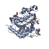
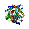
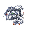
- PDB-4oua: Coexistent single-crystal structure of latent and active mushroom... -
+ Open data

- Basic information
| Entry | Database: PDB / ID: 4oua | ||||||
|---|---|---|---|---|---|---|---|
| Title | Coexistent single-crystal structure of latent and active mushroom tyrosinase (abPPO4) mediated by a hexatungstotellurate(VI) | ||||||
 Components Components |
| ||||||
 Keywords Keywords |  OXIDOREDUCTASE / type-3 copper protein / tyrosinase / PPO4 / zymogen / tyrosinase maturation / hetero-protein co-crystallization / Anderson-Evans-type polyoxometalate OXIDOREDUCTASE / type-3 copper protein / tyrosinase / PPO4 / zymogen / tyrosinase maturation / hetero-protein co-crystallization / Anderson-Evans-type polyoxometalate | ||||||
| Function / homology | di-copper center containing domain from catechol oxidase / Di-copper center containing domain from catechol oxidase / Orthogonal Bundle / Mainly Alpha / COPPER (I) ION / 6-tungstotellurate(VI) / :  Function and homology information Function and homology information | ||||||
| Biological species |  Agaricus bisporus var. bisporus H97 (fungus) Agaricus bisporus var. bisporus H97 (fungus) | ||||||
| Method | X-RAY DIFFRACTION / SYNCHROTRON / Resolution: 2.763 Å | ||||||
 Authors Authors | St.Mauracher, G. / Molitor, C. / Al-Oweini, R. / Kortz, U. / Rompel, A. | ||||||
 Citation Citation | Journal: Acta Crystallogr.,Sect.D / Year: 2014 Title: Latent and active abPPO4 mushroom tyrosinase cocrystallized with hexatungstotellurate(VI) in a single crystal. Authors: Mauracher, S.G. / Molitor, C. / Al-Oweini, R. / Kortz, U. / Rompel, A. #1: Journal: Acta Crystallogr.,Sect.F / Year: 2014 Title: Crystallization and preliminary X-ray crystallographic analysis of latent isoform PPO4 mushroom (Agaricus bisporus) tyrosinase. Authors: Mauracher, S.G. / Molitor, C. / Al-Oweini, R. / Kortz, U. / Rompel, A. #2: Journal: Phytochemistry / Year: 2014 Title: High level protein-purification allows the unambiguous polypeptide determination of latent isoform PPO4 of mushroom tyrosinase. Authors: Mauracher, S.G. / Molitor, C. / Michael, C. / Kragl, M. / Rizzi, A. / Rompel, A. | ||||||
| History |
|
- Structure visualization
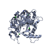
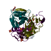
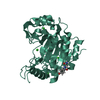
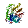
| Structure viewer | Molecule: MolmilJmol/JSmol |
|---|
- Downloads & links
-Download
| PDBx/mmCIF format | 4oua.cif.gz | 200.8 KB | Display | PDBx/mmCIF format |
|---|---|---|---|---|
| PDB format | pdb4oua.ent.gz | 166.8 KB | Display | PDB format |
| PDBx/mmJSON format | 4oua.json.gz | Tree view | PDBx/mmJSON format | |
| Others |  Other downloads Other downloads |
-Validation report
| Arichive directory | https://data.pdbj.org/pub/pdb/validation_reports/ou/4ouaftp://data.pdbj.org/pub/pdb/validation_reports/ou/4oua | HTTPS FTP |
|---|
-Related structure data
| Similar structure data |
|---|


-Links
 PDBj
PDBj- Assembly
| Deposited unit | 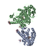
| |||||||||
|---|---|---|---|---|---|---|---|---|---|---|
| 1 | 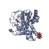
| |||||||||
| 2 | 
| |||||||||
| Unit cell |
| |||||||||
| Components on special symmetry positions |
|
-Components
-Protein , 2 types, 2 molecules AB
| #1: Protein | Mass: 43731.812 Da / Num. of mol.: 1 / Source method: isolated from a natural source / Details: White edible mushroom Source: (natural) Agaricus bisporus var. bisporus H97 (fungus)Strain: strain H97 / ATCC MYA-4626 / FGSC 10389 / References: UniProt: K9I869, tyrosinase |
|---|---|
| #2: Protein | Mass: 63639.168 Da / Num. of mol.: 1 / Source method: isolated from a natural source / Details: White edible mushroom Source: (natural) Agaricus bisporus var. bisporus H97 (fungus)Strain: strain H97 / ATCC MYA-4626 / FGSC 10389 / References: UniProt: K9I869, tyrosinase |
-Non-polymers , 5 types, 143 molecules 


| #3: Chemical | ChemComp-CU1 / Copper Mass: 63.546 Da / Num. of mol.: 4 / Source method: obtained synthetically / Formula: Cu Mass: 63.546 Da / Num. of mol.: 4 / Source method: obtained synthetically / Formula: Cu#4: Chemical | ChemComp-NA /  Mass: 22.990 Da / Num. of mol.: 4 / Source method: obtained synthetically / Formula: Na Mass: 22.990 Da / Num. of mol.: 4 / Source method: obtained synthetically / Formula: Na#5: Chemical | ChemComp-TRS / Tris Mass: 122.143 Da / Num. of mol.: 4 / Source method: obtained synthetically / Formula: C4H12NO3 / Comment: pH buffer*YM Mass: 122.143 Da / Num. of mol.: 4 / Source method: obtained synthetically / Formula: C4H12NO3 / Comment: pH buffer*YM#6: Chemical |  Mass: 1614.626 Da / Num. of mol.: 2 / Source method: obtained synthetically / Formula: O24TeW6 Mass: 1614.626 Da / Num. of mol.: 2 / Source method: obtained synthetically / Formula: O24TeW6#7: Water | ChemComp-HOH / | WaterMass: 18.015 Da / Num. of mol.: 129 / Source method: isolated from a natural source / Formula: H2O |
|---|
-Experimental details
-Experiment
| Experiment | Method: X-RAY DIFFRACTION / Number of used crystals: 1 |
|---|
- Sample preparation
| Crystal | Density Matthews: 2.72 Å3/Da / Density % sol: 54.78 % |
|---|---|
| Crystal grow | Temperature: 291 K / Method: vapor diffusion, hanging drop / pH: 7.5 Details: 10% PEG4000, 1 mM Na6[TeW6O24].22H2O, 25 mM Tris-HCl, pH 7.5, VAPOR DIFFUSION, HANGING DROP, temperature 291K |
-Data collection
| Diffraction | Mean temperature: 100 K | ||||||||||||||||||||||||||||||||||||||||||||||||||||||||||||||||||||||||||||||||||||||||||||||||||||||||||||||||||||||||||||||||||||||||||||
|---|---|---|---|---|---|---|---|---|---|---|---|---|---|---|---|---|---|---|---|---|---|---|---|---|---|---|---|---|---|---|---|---|---|---|---|---|---|---|---|---|---|---|---|---|---|---|---|---|---|---|---|---|---|---|---|---|---|---|---|---|---|---|---|---|---|---|---|---|---|---|---|---|---|---|---|---|---|---|---|---|---|---|---|---|---|---|---|---|---|---|---|---|---|---|---|---|---|---|---|---|---|---|---|---|---|---|---|---|---|---|---|---|---|---|---|---|---|---|---|---|---|---|---|---|---|---|---|---|---|---|---|---|---|---|---|---|---|---|---|---|---|
| Diffraction source | Source: SYNCHROTRON / Site: Diamond  / Beamline: I04-1 / Wavelength: 0.9173 Å / Beamline: I04-1 / Wavelength: 0.9173 Å | ||||||||||||||||||||||||||||||||||||||||||||||||||||||||||||||||||||||||||||||||||||||||||||||||||||||||||||||||||||||||||||||||||||||||||||
| Detector | Type: PSI PILATUS 6M / Detector: PIXEL / Date: May 15, 2013 | ||||||||||||||||||||||||||||||||||||||||||||||||||||||||||||||||||||||||||||||||||||||||||||||||||||||||||||||||||||||||||||||||||||||||||||
| Radiation | Monochromator: single bounce monochromator (22m) / Protocol: SINGLE WAVELENGTH / Monochromatic (M) / Laue (L): M / Scattering type: x-ray | ||||||||||||||||||||||||||||||||||||||||||||||||||||||||||||||||||||||||||||||||||||||||||||||||||||||||||||||||||||||||||||||||||||||||||||
| Radiation wavelength | Wavelength: 0.9173 Å / Relative weight: 1 | ||||||||||||||||||||||||||||||||||||||||||||||||||||||||||||||||||||||||||||||||||||||||||||||||||||||||||||||||||||||||||||||||||||||||||||
| Reflection | Resolution: 2.76→48.132 Å / Num. all: 28294 / % possible obs: 92.6 % / Observed criterion σ(F): 2 / Observed criterion σ(I): 2 / Biso Wilson estimate: 44.95 Å2 / Rmerge(I) obs: 0.129 / Χ2: 1.206 / Net I/σ(I): 6.53 | ||||||||||||||||||||||||||||||||||||||||||||||||||||||||||||||||||||||||||||||||||||||||||||||||||||||||||||||||||||||||||||||||||||||||||||
| Reflection shell |
|
- Processing
| Software |
| |||||||||||||||||||||||||||||||||||||||||||||||||||||||||||||||||||||||||||||
|---|---|---|---|---|---|---|---|---|---|---|---|---|---|---|---|---|---|---|---|---|---|---|---|---|---|---|---|---|---|---|---|---|---|---|---|---|---|---|---|---|---|---|---|---|---|---|---|---|---|---|---|---|---|---|---|---|---|---|---|---|---|---|---|---|---|---|---|---|---|---|---|---|---|---|---|---|---|---|
| Refinement | Resolution: 2.763→48.132 Å / FOM work R set: 0.8407 / SU ML: 0.37 / σ(F): 1.35 / Phase error: 23.36 / Stereochemistry target values: ML
| |||||||||||||||||||||||||||||||||||||||||||||||||||||||||||||||||||||||||||||
| Solvent computation | Shrinkage radii: 0.9 Å / VDW probe radii: 1.11 Å / Solvent model: FLAT BULK SOLVENT MODEL | |||||||||||||||||||||||||||||||||||||||||||||||||||||||||||||||||||||||||||||
| Displacement parameters | Biso max: 120.13 Å2 / Biso mean: 42.93 Å2 / Biso min: 18.88 Å2 | |||||||||||||||||||||||||||||||||||||||||||||||||||||||||||||||||||||||||||||
| Refinement step | Cycle: LAST / Resolution: 2.763→48.132 Å
| |||||||||||||||||||||||||||||||||||||||||||||||||||||||||||||||||||||||||||||
| Refine LS restraints |
| |||||||||||||||||||||||||||||||||||||||||||||||||||||||||||||||||||||||||||||
| LS refinement shell | Refine-ID: X-RAY DIFFRACTION / Total num. of bins used: 10
|
