 Movie
Movie Controller


+ Open data

- Basic information
| Entry | Database: PDB / ID: 5j45 | ||||||
|---|---|---|---|---|---|---|---|
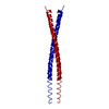
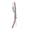
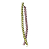
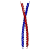
| Title | Crystal structure of Shrub, fly ortholog of SNF7/CHMP4B | ||||||
 Components Components | GH13992p | ||||||
 Keywords Keywords |  TRANSPORT PROTEIN / ESCRT / polymerization / membrane TRANSPORT PROTEIN / ESCRT / polymerization / membrane | ||||||
| Function / homology |  Function and homology information Function and homology informationEndosomal Sorting Complex Required For Transport (ESCRT) / Sealing of the nuclear envelope (NE) by ESCRT-III / fusome / Macroautophagy / contractile ring / female germ-line stem cell asymmetric division / ESCRT III complex / proximal dendrite / endosome transport via multivesicular body sorting pathway / late endosome to vacuole transport via multivesicular body sorting pathway ...Endosomal Sorting Complex Required For Transport (ESCRT) / Sealing of the nuclear envelope (NE) by ESCRT-III / fusome / Macroautophagy / contractile ring / female germ-line stem cell asymmetric division / ESCRT III complex / proximal dendrite / endosome transport via multivesicular body sorting pathway / late endosome to vacuole transport via multivesicular body sorting pathway / vesicle budding from membrane / ubiquitin-dependent protein catabolic process via the multivesicular body sorting pathway / dendrite morphogenesis / neuron remodeling / mitotic cytokinesis / multivesicular body / cytoplasmic side of plasma membrane / autophagy / midbody / symbiont entry into host cell / neuronal cell bodySimilarity search - Function | ||||||
| Biological species |  Drosophila melanogaster (fruit fly) Drosophila melanogaster (fruit fly) | ||||||
| Method | X-RAY DIFFRACTION / SYNCHROTRON / Resolution: 2.758 Å | ||||||
 Authors Authors | McMillan, B.J. / Blacklow, S.C. | ||||||
 Citation Citation | Journal: Cell Rep / Year: 2016 Title: Electrostatic Interactions between Elongated Monomers Drive Filamentation of Drosophila Shrub, a Metazoan ESCRT-III Protein. Authors: McMillan, B.J. / Tibbe, C. / Jeon, H. / Drabek, A.A. / Klein, T. / Blacklow, S.C. | ||||||
| History |
|
- Structure visualization
| Structure viewer | Molecule: MolmilJmol/JSmol |
|---|
- Downloads & links
-Download
| PDBx/mmCIF format | 5j45.cif.gz | 75.8 KB | Display | PDBx/mmCIF format |
|---|---|---|---|---|
| PDB format | pdb5j45.ent.gz | 63 KB | Display | PDB format |
| PDBx/mmJSON format | 5j45.json.gz | Tree view | PDBx/mmJSON format | |
| Others |  Other downloads Other downloads |
-Validation report
| Arichive directory | https://data.pdbj.org/pub/pdb/validation_reports/j4/5j45ftp://data.pdbj.org/pub/pdb/validation_reports/j4/5j45 | HTTPS FTP |
|---|
-Related structure data
| Similar structure data |
|---|


-Links
 PDBj
PDBj- Assembly
| Deposited unit | 
| ||||||||
|---|---|---|---|---|---|---|---|---|---|
| 1 |
| ||||||||
| Unit cell |
|
-Components
| #1: Protein | Mass: 14754.847 Da / Num. of mol.: 1 / Fragment: UNP residues 18-143 Source method: isolated from a genetically manipulated source Source: (gene. exp.) Drosophila melanogaster (fruit fly) / Gene: shrb, Vps32, CG8055, Dmel_CG8055 / Production host:  Escherichia coli (E. coli) / Variant (production host): BL21 PLysS / References: UniProt: Q8T0Q4 Escherichia coli (E. coli) / Variant (production host): BL21 PLysS / References: UniProt: Q8T0Q4 |
|---|
-Experimental details
-Experiment
| Experiment | Method: X-RAY DIFFRACTION / Number of used crystals: 1 |
|---|
- Sample preparation
| Crystal | Density Matthews: 2.67 Å3/Da / Density % sol: 53.9 % |
|---|---|
| Crystal grow | Temperature: 293.15 K / Method: vapor diffusion, hanging drop / pH: 5.5 Details: 17% v/v PEG10K, 0.1 M NH4CH3CO2, and 100 mM Bis-Tris pH 5.5 |
-Data collection
| Diffraction | Mean temperature: 100 K |
|---|---|
| Diffraction source | Source: SYNCHROTRON / Site: APS  / Beamline: 24-ID-C / Wavelength: 0.9792 Å / Beamline: 24-ID-C / Wavelength: 0.9792 Å |
| Detector | Type: DECTRIS PILATUS 6M-F / Detector: PIXEL / Date: Mar 19, 2015 |
| Radiation | Protocol: SINGLE WAVELENGTH / Monochromatic (M) / Laue (L): M / Scattering type: x-ray |
| Radiation wavelength | Wavelength: 0.9792 Å / Relative weight: 1 |
| Reflection | Resolution: 2.758→50 Å / Num. obs: 3869 / % possible obs: 98.1 % / Redundancy: 3.1 % / Biso Wilson estimate: 85.75 Å2 / Rmerge(I) obs: 0.09 / Net I/σ(I): 15.9 |
| Reflection shell | Highest resolution: 2.758 Å |
- Processing
| Software |
| ||||||||||||||||||||||||||||||||||||||||||||||||||||||||||||||||||||||||||||||||||||||||||||||||||||||||||||||||||||||||||||||||||||||||||||||||||||||
|---|---|---|---|---|---|---|---|---|---|---|---|---|---|---|---|---|---|---|---|---|---|---|---|---|---|---|---|---|---|---|---|---|---|---|---|---|---|---|---|---|---|---|---|---|---|---|---|---|---|---|---|---|---|---|---|---|---|---|---|---|---|---|---|---|---|---|---|---|---|---|---|---|---|---|---|---|---|---|---|---|---|---|---|---|---|---|---|---|---|---|---|---|---|---|---|---|---|---|---|---|---|---|---|---|---|---|---|---|---|---|---|---|---|---|---|---|---|---|---|---|---|---|---|---|---|---|---|---|---|---|---|---|---|---|---|---|---|---|---|---|---|---|---|---|---|---|---|---|---|---|---|
| Refinement | Resolution: 2.758→43.578 Å / SU ML: 0.46 / Cross valid method: FREE R-VALUE / σ(F): 0 / Phase error: 33.76
| ||||||||||||||||||||||||||||||||||||||||||||||||||||||||||||||||||||||||||||||||||||||||||||||||||||||||||||||||||||||||||||||||||||||||||||||||||||||
| Solvent computation | Shrinkage radii: 0.9 Å / VDW probe radii: 1.11 Å | ||||||||||||||||||||||||||||||||||||||||||||||||||||||||||||||||||||||||||||||||||||||||||||||||||||||||||||||||||||||||||||||||||||||||||||||||||||||
| Refinement step | Cycle: LAST / Resolution: 2.758→43.578 Å
| ||||||||||||||||||||||||||||||||||||||||||||||||||||||||||||||||||||||||||||||||||||||||||||||||||||||||||||||||||||||||||||||||||||||||||||||||||||||
| Refine LS restraints |
| ||||||||||||||||||||||||||||||||||||||||||||||||||||||||||||||||||||||||||||||||||||||||||||||||||||||||||||||||||||||||||||||||||||||||||||||||||||||
| LS refinement shell |
| ||||||||||||||||||||||||||||||||||||||||||||||||||||||||||||||||||||||||||||||||||||||||||||||||||||||||||||||||||||||||||||||||||||||||||||||||||||||
| Refinement TLS params. | Method: refined / Refine-ID: X-RAY DIFFRACTION
| ||||||||||||||||||||||||||||||||||||||||||||||||||||||||||||||||||||||||||||||||||||||||||||||||||||||||||||||||||||||||||||||||||||||||||||||||||||||
| Refinement TLS group |
|
