 Movie
Movie Controller

[English] 日本語
 Yorodumi
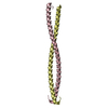
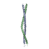
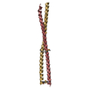
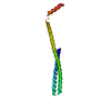
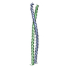
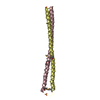
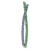
- PDB-4h22: Crystal structure of the dimeric coiled-coil domain of the cytoso... -
+ Open data

- Basic information
| Entry | Database: PDB / ID: 4h22 | ||||||
|---|---|---|---|---|---|---|---|
| Title | Crystal structure of the dimeric coiled-coil domain of the cytosolic nucleic acid sensor LRRFIP1 | ||||||
 Components Components | Leucine-rich repeat flightless-interacting protein 1 | ||||||
 Keywords Keywords |  TRANSCRIPTION / nucleic acid sensor / Flightless-1 TRANSCRIPTION / nucleic acid sensor / Flightless-1 | ||||||
| Function / homology |  Function and homology information Function and homology informationLRR FLII-interacting protein 1 (LRRFIP1) activates type I IFN production / Signaling by cytosolic FGFR1 fusion mutants / positive regulation of type I interferon production / Signaling by FGFR1 in disease / DNA-binding transcription repressor activity, RNA polymerase II-specific / double-stranded RNA binding / positive regulation of NF-kappaB transcription factor activity / cytoskeleton / cadherin binding / RNA polymerase II cis-regulatory region sequence-specific DNA binding ...LRR FLII-interacting protein 1 (LRRFIP1) activates type I IFN production / Signaling by cytosolic FGFR1 fusion mutants / positive regulation of type I interferon production / Signaling by FGFR1 in disease / DNA-binding transcription repressor activity, RNA polymerase II-specific / double-stranded RNA binding / positive regulation of NF-kappaB transcription factor activity / cytoskeleton / cadherin binding / RNA polymerase II cis-regulatory region sequence-specific DNA binding / negative regulation of DNA-templated transcription / regulation of transcription by RNA polymerase II / negative regulation of transcription by RNA polymerase II / protein homodimerization activity / DNA binding / nucleus / plasma membrane / cytosol / cytoplasmSimilarity search - Function | ||||||
| Biological species |  Homo sapiens (human) Homo sapiens (human) | ||||||
| Method | X-RAY DIFFRACTION / SYNCHROTRON / MOLECULAR REPLACEMENT / Resolution: 2.89 Å | ||||||
 Authors Authors | Nguyen, J.B. / Modis, Y. | ||||||
 Citation Citation | Journal: J.Struct.Biol. / Year: 2013 Title: Crystal structure of the dimeric coiled-coil domain of the cytosolic nucleic acid sensor LRRFIP1. Authors: Nguyen, J.B. / Modis, Y. | ||||||
| History |
|
- Structure visualization
| Structure viewer | Molecule: MolmilJmol/JSmol |
|---|
- Downloads & links
-Download
| PDBx/mmCIF format | 4h22.cif.gz | 142.6 KB | Display | PDBx/mmCIF format |
|---|---|---|---|---|
| PDB format | pdb4h22.ent.gz | 115.4 KB | Display | PDB format |
| PDBx/mmJSON format | 4h22.json.gz | Tree view | PDBx/mmJSON format | |
| Others |  Other downloads Other downloads |
-Validation report
| Arichive directory | https://data.pdbj.org/pub/pdb/validation_reports/h2/4h22ftp://data.pdbj.org/pub/pdb/validation_reports/h2/4h22 | HTTPS FTP |
|---|
-Related structure data
| Related structure data |  2q6qS S: Starting model for refinement |
|---|---|
| Similar structure data |


-Links
 PDBj
PDBj


- Assembly
| Deposited unit | 
| ||||||||
|---|---|---|---|---|---|---|---|---|---|
| 1 | 
| ||||||||
| 2 | 
| ||||||||
| Unit cell |
|
-Components
| #1: Protein | Mass: 12399.946 Da / Num. of mol.: 4 / Fragment: UNP residues 162-249 Source method: isolated from a genetically manipulated source Source: (gene. exp.) Homo sapiens (human) / Gene: LRRFIP1, GCF2, TRIP / Plasmid: pET21a / Production host:  Escherichia coli (E. coli) / Strain (production host): Rosetta / References: UniProt: Q32MZ4 Escherichia coli (E. coli) / Strain (production host): Rosetta / References: UniProt: Q32MZ4#2: Water | ChemComp-HOH / | Water Mass: 18.015 Da / Num. of mol.: 26 / Source method: isolated from a natural source / Formula: H2O Mass: 18.015 Da / Num. of mol.: 26 / Source method: isolated from a natural source / Formula: H2O |
|---|
-Experimental details
-Experiment
| Experiment | Method: X-RAY DIFFRACTION / Number of used crystals: 1 |
|---|
- Sample preparation
| Crystal | Density Matthews: 3.51 Å3/Da / Density % sol: 64.91 % |
|---|---|
| Crystal grow | Temperature: 289 K / Method: vapor diffusion, hanging drop / pH: 6.5 Details: PEG 8000, MES pH 6.5, sodium acetate, VAPOR DIFFUSION, HANGING DROP, temperature 289K |
-Data collection
| Diffraction | Mean temperature: 100 K |
|---|---|
| Diffraction source | Source: SYNCHROTRON / Site: NSLS  / Beamline: X29A / Wavelength: 1.075 Å / Beamline: X29A / Wavelength: 1.075 Å |
| Detector | Type: ADSC QUANTUM 315r / Detector: CCD / Date: Nov 4, 2010 |
| Radiation | Monochromator: Si(111) / Protocol: SINGLE WAVELENGTH / Monochromatic (M) / Laue (L): M / Scattering type: x-ray |
| Radiation wavelength | Wavelength: 1.075 Å / Relative weight: 1 |
| Reflection | Resolution: 2.89→50 Å / Num. all: 15508 / Num. obs: 15465 / % possible obs: 99.7 % / Observed criterion σ(F): 1.8 / Observed criterion σ(I): 1.8 |
| Reflection shell | Resolution: 2.89→2.94 Å / % possible all: 95.5 |
- Processing
| Software |
| |||||||||||||||||||||||||||||||||||||||||||||||||||||||||||||||||||||||||||||||||||||||||||||||||||||||||||||||||||||||||||||
|---|---|---|---|---|---|---|---|---|---|---|---|---|---|---|---|---|---|---|---|---|---|---|---|---|---|---|---|---|---|---|---|---|---|---|---|---|---|---|---|---|---|---|---|---|---|---|---|---|---|---|---|---|---|---|---|---|---|---|---|---|---|---|---|---|---|---|---|---|---|---|---|---|---|---|---|---|---|---|---|---|---|---|---|---|---|---|---|---|---|---|---|---|---|---|---|---|---|---|---|---|---|---|---|---|---|---|---|---|---|---|---|---|---|---|---|---|---|---|---|---|---|---|---|---|---|---|
| Refinement | Method to determine structure: MOLECULAR REPLACEMENT Starting model: PDB entry 2q6q Resolution: 2.89→50 Å / Cor.coef. Fo:Fc: 0.934 / Cor.coef. Fo:Fc free: 0.923 / SU B: 29.59 / SU ML: 0.255 / Cross valid method: THROUGHOUT / σ(F): 1.8 / ESU R: 0.434 / ESU R Free: 0.31 / Stereochemistry target values: MAXIMUM LIKELIHOOD / Details: HYDROGENS HAVE BEEN USED IF PRESENT IN THE INPUT
| |||||||||||||||||||||||||||||||||||||||||||||||||||||||||||||||||||||||||||||||||||||||||||||||||||||||||||||||||||||||||||||
| Solvent computation | Ion probe radii: 0.8 Å / Shrinkage radii: 0.8 Å / VDW probe radii: 1.2 Å / Solvent model: MASK | |||||||||||||||||||||||||||||||||||||||||||||||||||||||||||||||||||||||||||||||||||||||||||||||||||||||||||||||||||||||||||||
| Displacement parameters | Biso mean: 100.862 Å2
| |||||||||||||||||||||||||||||||||||||||||||||||||||||||||||||||||||||||||||||||||||||||||||||||||||||||||||||||||||||||||||||
| Refinement step | Cycle: LAST / Resolution: 2.89→50 Å
| |||||||||||||||||||||||||||||||||||||||||||||||||||||||||||||||||||||||||||||||||||||||||||||||||||||||||||||||||||||||||||||
| Refine LS restraints |
| |||||||||||||||||||||||||||||||||||||||||||||||||||||||||||||||||||||||||||||||||||||||||||||||||||||||||||||||||||||||||||||
| LS refinement shell | Resolution: 2.89→2.962 Å / Total num. of bins used: 20
| |||||||||||||||||||||||||||||||||||||||||||||||||||||||||||||||||||||||||||||||||||||||||||||||||||||||||||||||||||||||||||||
| Refinement TLS params. | Method: refined / Refine-ID: X-RAY DIFFRACTION
| |||||||||||||||||||||||||||||||||||||||||||||||||||||||||||||||||||||||||||||||||||||||||||||||||||||||||||||||||||||||||||||
| Refinement TLS group |
|
