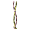
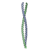
 Movie
Movie Controller

[English] 日本語
 Yorodumi
- PDB-5fd7: X-ray Crystal Structure of ESCRT-III Snf7 core domain (conformation A) -
+ Open data

- Basic information
| Entry | Database: PDB / ID: 5fd7 | |||||||||||||||
|---|---|---|---|---|---|---|---|---|---|---|---|---|---|---|---|---|
| Title | X-ray Crystal Structure of ESCRT-III Snf7 core domain (conformation A) | |||||||||||||||
 Components Components | Vacuolar-sorting protein SNF7 | |||||||||||||||
 Keywords Keywords |  PROTEIN TRANSPORT / ESCRT / Snf7 / Active / Core PROTEIN TRANSPORT / ESCRT / Snf7 / Active / Core | |||||||||||||||
| Function / homology |  Function and homology information Function and homology informationESCRT III complex assembly / intralumenal vesicle formation / Sealing of the nuclear envelope (NE) by ESCRT-III / Macroautophagy / ATP export / ESCRT III complex / Endosomal Sorting Complex Required For Transport (ESCRT) / late endosome to vacuole transport via multivesicular body sorting pathway / vesicle budding from membrane / late endosome to vacuole transport ...ESCRT III complex assembly / intralumenal vesicle formation / Sealing of the nuclear envelope (NE) by ESCRT-III / Macroautophagy / ATP export / ESCRT III complex / Endosomal Sorting Complex Required For Transport (ESCRT) / late endosome to vacuole transport via multivesicular body sorting pathway / vesicle budding from membrane / late endosome to vacuole transport / ubiquitin-dependent protein catabolic process via the multivesicular body sorting pathway / reticulophagy / multivesicular body / cytoplasmic side of plasma membrane / protein transport / nuclear envelope / identical protein binding / plasma membrane / cytoplasmSimilarity search - Function | |||||||||||||||
| Biological species |  Saccharomyces cerevisiae (brewer's yeast) Saccharomyces cerevisiae (brewer's yeast) | |||||||||||||||
| Method | X-RAY DIFFRACTION / SYNCHROTRON / MOLECULAR REPLACEMENT / molecular replacement / Resolution: 2.4 Å | |||||||||||||||
 Authors Authors | Tang, S. / Henne, W.M. / Borbat, P.P. / Buchkovich, N.J. / Freed, J.H. / Mao, Y. / Fromme, J.C. / Emr, S.D. | |||||||||||||||
| Funding support |  United States, 4items United States, 4items
| |||||||||||||||
 Citation Citation | Journal: Elife / Year: 2015 Title: Structural basis for activation, assembly and membrane binding of ESCRT-III Snf7 filaments. Authors: Tang, S. / Henne, W.M. / Borbat, P.P. / Buchkovich, N.J. / Freed, J.H. / Mao, Y. / Fromme, J.C. / Emr, S.D. | |||||||||||||||
| History |
|
- Structure visualization
| Structure viewer | Molecule: MolmilJmol/JSmol |
|---|
- Downloads & links
-Download
| PDBx/mmCIF format | 5fd7.cif.gz | 63.7 KB | Display | PDBx/mmCIF format |
|---|---|---|---|---|
| PDB format | pdb5fd7.ent.gz | 46.9 KB | Display | PDB format |
| PDBx/mmJSON format | 5fd7.json.gz | Tree view | PDBx/mmJSON format | |
| Others |  Other downloads Other downloads |
-Validation report
| Arichive directory | https://data.pdbj.org/pub/pdb/validation_reports/fd/5fd7ftp://data.pdbj.org/pub/pdb/validation_reports/fd/5fd7 | HTTPS FTP |
|---|
-Related structure data
| Related structure data | 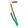 5fd9C 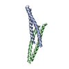 4abmS S: Starting model for refinement C: citing same article ( |
|---|---|
| Similar structure data |


-Links
 PDBj
PDBj

- Assembly
| Deposited unit | 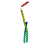
| ||||||||
|---|---|---|---|---|---|---|---|---|---|
| 1 |
| ||||||||
| Unit cell |
|
-Components
| #1: Protein | Mass: 15860.185 Da / Num. of mol.: 1 Source method: isolated from a genetically manipulated source Source: (gene. exp.) Saccharomyces cerevisiae (strain ATCC 204508 / S288c) (yeast)Strain: ATCC 204508 / S288c / Gene: SNF7, DID1, VPS32, YLR025W / Plasmid: pET28a His6-Sumo-Snf7N12-P150 / Production host:  Escherichia coli (E. coli) / Strain (production host): Rosetta / References: UniProt: P39929 Escherichia coli (E. coli) / Strain (production host): Rosetta / References: UniProt: P39929 |
|---|---|
| #2: Water | ChemComp-HOH / Water Mass: 18.015 Da / Num. of mol.: 7 / Source method: isolated from a natural source / Formula: H2O Mass: 18.015 Da / Num. of mol.: 7 / Source method: isolated from a natural source / Formula: H2O |
-Experimental details
-Experiment
| Experiment | Method: X-RAY DIFFRACTION / Number of used crystals: 1 |
|---|
- Sample preparation
| Crystal | Density Matthews: 2.63 Å3/Da / Density % sol: 53.18 % |
|---|---|
| Crystal grow | Temperature: 277 K / Method: vapor diffusion, hanging drop / pH: 5.5 / Details: 100mM NaCl, 100mM MES:NaOH pH5.5, 3% PEG20,000 |
-Data collection
| Diffraction | Mean temperature: 80 K |
|---|---|
| Diffraction source | Source: SYNCHROTRON / Site: CHESS / Beamline: F1 / Wavelength: 0.978 Å |
| Detector | Type: DECTRIS PILATUS 6M / Detector: PIXEL / Date: Mar 26, 2015 |
| Radiation | Protocol: SINGLE WAVELENGTH / Monochromatic (M) / Laue (L): M / Scattering type: x-ray |
| Radiation wavelength | Wavelength: 0.978 Å / Relative weight: 1 |
| Reflection | Resolution: 2.4→50 Å / Num. obs: 6376 / % possible obs: 97.99 % / Redundancy: 3.6 % / Rmerge(I) obs: 0.0884 / Net I/σ(I): 8.04 |
| Reflection shell | Resolution: 2.4→2.49 Å / Redundancy: 3.2 % / Rmerge(I) obs: 0.249 / Mean I/σ(I) obs: 2.91 / % possible all: 93.72 |
-Phasing
| Phasing | Method: molecular replacement |
|---|
- Processing
| Software |
| |||||||||||||||||||||||||||||||||||||||||||||||||||||||||||||||||||||||||||||||||||||||||||||||||||||||||||||||||||||||||||||
|---|---|---|---|---|---|---|---|---|---|---|---|---|---|---|---|---|---|---|---|---|---|---|---|---|---|---|---|---|---|---|---|---|---|---|---|---|---|---|---|---|---|---|---|---|---|---|---|---|---|---|---|---|---|---|---|---|---|---|---|---|---|---|---|---|---|---|---|---|---|---|---|---|---|---|---|---|---|---|---|---|---|---|---|---|---|---|---|---|---|---|---|---|---|---|---|---|---|---|---|---|---|---|---|---|---|---|---|---|---|---|---|---|---|---|---|---|---|---|---|---|---|---|---|---|---|---|
| Refinement | Method to determine structure: MOLECULAR REPLACEMENT Starting model: 4ABM Resolution: 2.4→27.009 Å / SU ML: 0.18 / Cross valid method: FREE R-VALUE / σ(F): 1.36 / Phase error: 38.56 / Stereochemistry target values: ML
| |||||||||||||||||||||||||||||||||||||||||||||||||||||||||||||||||||||||||||||||||||||||||||||||||||||||||||||||||||||||||||||
| Solvent computation | Shrinkage radii: 0.9 Å / VDW probe radii: 1.11 Å / Solvent model: FLAT BULK SOLVENT MODEL | |||||||||||||||||||||||||||||||||||||||||||||||||||||||||||||||||||||||||||||||||||||||||||||||||||||||||||||||||||||||||||||
| Displacement parameters | Biso max: 218.02 Å2 / Biso mean: 91.0538 Å2 / Biso min: 40.74 Å2 | |||||||||||||||||||||||||||||||||||||||||||||||||||||||||||||||||||||||||||||||||||||||||||||||||||||||||||||||||||||||||||||
| Refinement step | Cycle: final / Resolution: 2.4→27.009 Å
| |||||||||||||||||||||||||||||||||||||||||||||||||||||||||||||||||||||||||||||||||||||||||||||||||||||||||||||||||||||||||||||
| Refine LS restraints |
| |||||||||||||||||||||||||||||||||||||||||||||||||||||||||||||||||||||||||||||||||||||||||||||||||||||||||||||||||||||||||||||
| LS refinement shell | Refine-ID: X-RAY DIFFRACTION / Total num. of bins used: 2 / % reflection obs: 97 %
| |||||||||||||||||||||||||||||||||||||||||||||||||||||||||||||||||||||||||||||||||||||||||||||||||||||||||||||||||||||||||||||
| Refinement TLS params. | Method: refined / Refine-ID: X-RAY DIFFRACTION
| |||||||||||||||||||||||||||||||||||||||||||||||||||||||||||||||||||||||||||||||||||||||||||||||||||||||||||||||||||||||||||||
| Refinement TLS group |
|
