 Movie
Movie Controller

[English] 日本語
 Yorodumi
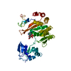
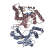
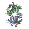
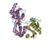
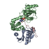
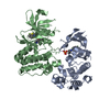
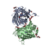
- PDB-5j3t: Crystal structure of S. pombe Dcp2:Dcp1:Edc1 mRNA decapping complex -
+ Open data

- Basic information
| Entry | Database: PDB / ID: 5j3t | ||||||
|---|---|---|---|---|---|---|---|
| Title | Crystal structure of S. pombe Dcp2:Dcp1:Edc1 mRNA decapping complex | ||||||
 Components Components |
| ||||||
 Keywords Keywords |  HYDROLASE / decapping / mRNA decay / EVH1 / Nudix HYDROLASE / decapping / mRNA decay / EVH1 / Nudix | ||||||
| Function / homology |  Function and homology information Function and homology informationmRNA phosphatase activator activity / : / RNA decapping complex / mRNA methylguanosine-cap decapping / 5'-(N(7)-methyl 5'-triphosphoguanosine)-[mRNA] diphosphatase activity / 5'-(N(7)-methylguanosine 5'-triphospho)-[mRNA] hydrolase activity / Hydrolases / deadenylation-dependent decapping of nuclear-transcribed mRNA / mRNA cap binding / : ...mRNA phosphatase activator activity / : / RNA decapping complex / mRNA methylguanosine-cap decapping / 5'-(N(7)-methyl 5'-triphosphoguanosine)-[mRNA] diphosphatase activity / 5'-(N(7)-methylguanosine 5'-triphospho)-[mRNA] hydrolase activity / Hydrolases / deadenylation-dependent decapping of nuclear-transcribed mRNA / mRNA cap binding / : / nuclear-transcribed mRNA catabolic process, nonsense-mediated decay / meiotic cell cycle / P-body / mRNA processing / cytoplasmic stress granule / manganese ion binding / single-stranded RNA binding / magnesium ion binding / RNA binding / ATP binding / nucleus / cytosol / cytoplasmSimilarity search - Function | ||||||
| Biological species |  Schizosaccharomyces pombe (fission yeast) Schizosaccharomyces pombe (fission yeast) | ||||||
| Method | X-RAY DIFFRACTION / SYNCHROTRON / MOLECULAR REPLACEMENT / Resolution: 1.6 Å | ||||||
 Authors Authors | Valkov, E. / Muthukumar, S. / Chang, C.T. / Jonas, S. / Weichenrieder, O. / Izaurralde, E. | ||||||
 Citation Citation | Journal: Nat.Struct.Mol.Biol. / Year: 2016 Title: Structure of the Dcp2-Dcp1 mRNA-decapping complex in the activated conformation. Authors: Valkov, E. / Muthukumar, S. / Chang, C.T. / Jonas, S. / Weichenrieder, O. / Izaurralde, E. | ||||||
| History |
| ||||||
| Remark 650 | HELIX DETERMINATION METHOD: AUTHOR PROVIDED. | ||||||
| Remark 700 | SHEET DETERMINATION METHOD: AUTHOR PROVIDED. |
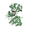
- Structure visualization
| Structure viewer | Molecule: MolmilJmol/JSmol |
|---|
- Downloads & links
-Download
| PDBx/mmCIF format | 5j3t.cif.gz | 179.7 KB | Display | PDBx/mmCIF format |
|---|---|---|---|---|
| PDB format | pdb5j3t.ent.gz | 142.3 KB | Display | PDB format |
| PDBx/mmJSON format | 5j3t.json.gz | Tree view | PDBx/mmJSON format | |
| Others |  Other downloads Other downloads |
-Validation report
| Arichive directory | https://data.pdbj.org/pub/pdb/validation_reports/j3/5j3tftp://data.pdbj.org/pub/pdb/validation_reports/j3/5j3t | HTTPS FTP |
|---|
-Related structure data
| Related structure data |  5j3qC  5j3ySC C: citing same article ( S: Starting model for refinement |
|---|---|
| Similar structure data |
-Links
 PDBj
PDBj

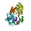
- Assembly
| Deposited unit | 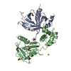
| |||||||||
|---|---|---|---|---|---|---|---|---|---|---|
| 1 |
| |||||||||
| Unit cell |
| |||||||||
| Components on special symmetry positions |
|
-Components
-Protein , 2 types, 2 molecules AB
| #1: Protein | Mass: 15294.453 Da / Num. of mol.: 1 Source method: isolated from a genetically manipulated source Source: (gene. exp.) Schizosaccharomyces pombe (fission yeast)Gene: dcp1, SPBC3B9.21 / Plasmid: pnEApG / Production host:  Escherichia coli BL21(DE3) (bacteria) / Variant (production host): STar / References: UniProt: Q9P805 Escherichia coli BL21(DE3) (bacteria) / Variant (production host): STar / References: UniProt: Q9P805 |
|---|---|
| #2: Protein | Mass: 28399.793 Da / Num. of mol.: 1 Source method: isolated from a genetically manipulated source Source: (gene. exp.) Schizosaccharomyces pombe (fission yeast)Strain: 972 / ATCC 24843 / Gene: dcp2, SPAC19A8.12 / Plasmid: pnYC / Production host: Escherichia coli BL21(DE3) (bacteria) / Variant (production host): StarReferences: UniProt: O13828, 5'-(N7-methylguanosine 5'-triphospho)-[mRNA] hydrolase |
-Protein/peptide , 1 types, 1 molecules C
| #3: Protein/peptide | Mass: 2742.089 Da / Num. of mol.: 1 / Source method: obtained synthetically / Source: (synth.) Schizosaccharomyces pombe (fission yeast) / References: UniProt: Q10108 |
|---|
-Non-polymers , 3 types, 221 molecules 


| #4: Chemical | ChemComp-FMT / Formic acid Mass: 46.025 Da / Num. of mol.: 5 / Source method: obtained synthetically / Formula: CH2O2 Mass: 46.025 Da / Num. of mol.: 5 / Source method: obtained synthetically / Formula: CH2O2#5: Chemical | ChemComp-MG / |  Mass: 24.305 Da / Num. of mol.: 1 / Source method: obtained synthetically / Formula: Mg Mass: 24.305 Da / Num. of mol.: 1 / Source method: obtained synthetically / Formula: Mg#6: Water | ChemComp-HOH / | WaterMass: 18.015 Da / Num. of mol.: 215 / Source method: isolated from a natural source / Formula: H2O |
|---|
-Experimental details
-Experiment
| Experiment | Method: X-RAY DIFFRACTION / Number of used crystals: 1 |
|---|
- Sample preparation
| Crystal | Density Matthews: 1.9 Å3/Da / Density % sol: 35 % |
|---|---|
| Crystal grow | Temperature: 292 K / Method: vapor diffusion / pH: 4.8 Details: 1.55 M sodium formate, 0.1 M sodium acetate (pH 4.8) |
-Data collection
| Diffraction | Mean temperature: 100 K |
|---|---|
| Diffraction source | Source: SYNCHROTRON / Site: SLS  / Beamline: X10SA / Wavelength: 1 Å / Beamline: X10SA / Wavelength: 1 Å |
| Detector | Type: DECTRIS PILATUS 6M-F / Detector: PIXEL / Date: Apr 19, 2015 / Details: DYNAMICALLY BENDABLE MIRRORS |
| Radiation | Monochromator: Si(111) / Protocol: SINGLE WAVELENGTH / Monochromatic (M) / Laue (L): M / Scattering type: x-ray |
| Radiation wavelength | Wavelength: 1 Å / Relative weight: 1 |
| Reflection | Resolution: 1.6→45.1 Å / Num. obs: 45926 / % possible obs: 99.1 % / Redundancy: 3.2 % / Biso Wilson estimate: 31.59 Å2 / Rsym value: 0.028 / Net I/σ(I): 18.9 |
| Reflection shell | Resolution: 1.6→1.68 Å / Redundancy: 3.2 % / Mean I/σ(I) obs: 1.6 / Rsym value: 0.653 / % possible all: 96.4 |
- Processing
| Software |
| ||||||||||||||||||||||||||||||||||||||||||||||||||||||||||||||||||||||||||||||||||||||||||||||||||||||||||||||||||
|---|---|---|---|---|---|---|---|---|---|---|---|---|---|---|---|---|---|---|---|---|---|---|---|---|---|---|---|---|---|---|---|---|---|---|---|---|---|---|---|---|---|---|---|---|---|---|---|---|---|---|---|---|---|---|---|---|---|---|---|---|---|---|---|---|---|---|---|---|---|---|---|---|---|---|---|---|---|---|---|---|---|---|---|---|---|---|---|---|---|---|---|---|---|---|---|---|---|---|---|---|---|---|---|---|---|---|---|---|---|---|---|---|---|---|---|
| Refinement | Method to determine structure: MOLECULAR REPLACEMENT Starting model: 5J3Y Resolution: 1.6→23.43 Å / Cor.coef. Fo:Fc: 0.965 / Cor.coef. Fo:Fc free: 0.9505 / SU R Cruickshank DPI: 0.098 / Cross valid method: THROUGHOUT / σ(F): 0 / SU R Blow DPI: 0.099 / SU Rfree Blow DPI: 0.096 / SU Rfree Cruickshank DPI: 0.096
| ||||||||||||||||||||||||||||||||||||||||||||||||||||||||||||||||||||||||||||||||||||||||||||||||||||||||||||||||||
| Displacement parameters | Biso mean: 40.78 Å2
| ||||||||||||||||||||||||||||||||||||||||||||||||||||||||||||||||||||||||||||||||||||||||||||||||||||||||||||||||||
| Refine analyze | Luzzati coordinate error obs: 0.216 Å | ||||||||||||||||||||||||||||||||||||||||||||||||||||||||||||||||||||||||||||||||||||||||||||||||||||||||||||||||||
| Refinement step | Cycle: LAST / Resolution: 1.6→23.43 Å
| ||||||||||||||||||||||||||||||||||||||||||||||||||||||||||||||||||||||||||||||||||||||||||||||||||||||||||||||||||
| Refine LS restraints |
| ||||||||||||||||||||||||||||||||||||||||||||||||||||||||||||||||||||||||||||||||||||||||||||||||||||||||||||||||||
| LS refinement shell | Resolution: 1.6→1.64 Å / Total num. of bins used: 20
| ||||||||||||||||||||||||||||||||||||||||||||||||||||||||||||||||||||||||||||||||||||||||||||||||||||||||||||||||||
| Refinement TLS params. | Method: refined / Refine-ID: X-RAY DIFFRACTION
| ||||||||||||||||||||||||||||||||||||||||||||||||||||||||||||||||||||||||||||||||||||||||||||||||||||||||||||||||||
| Refinement TLS group |
|
