 Movie
Movie Controller


+ Open data

- Basic information
| Entry | Database: PDB / ID: 3tpa | ||||||
|---|---|---|---|---|---|---|---|
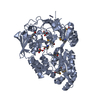
| Title | Structure of HbpA2 from Haemophilus parasuis | ||||||
 Components Components | Heme-binding protein A | ||||||
 Keywords Keywords | HEME BINDING PROTEIN / Glutathione binding protein / SBP | ||||||
| Function / homology |  Function and homology information Function and homology informationDipeptide-binding Protein; domain 1 / Dipeptide-binding Protein; Domain 1 / Dipeptide-binding Protein; domain 3 / Dipeptide-binding Protein; Domain 3 / Periplasmic binding protein-like II / D-Maltodextrin-Binding Protein; domain 2 / Roll /  Alpha-Beta Complex / 3-Layer(aba) Sandwich / Alpha Beta Alpha-Beta Complex / 3-Layer(aba) Sandwich / Alpha BetaSimilarity search - Domain/homology | ||||||
| Biological species |  Haemophilus parasuis 29755 (bacteria) Haemophilus parasuis 29755 (bacteria) | ||||||
| Method | X-RAY DIFFRACTION / SYNCHROTRON / MOLECULAR REPLACEMENT / Resolution: 2.0001 Å | ||||||
 Authors Authors | Vergauwen, B. / Van der Meeren, R. / Dansercoer, A. / Savvides, S.N. | ||||||
 Citation Citation | Journal: Bmc Biochem. / Year: 2011 Title: Delineation of the Pasteurellaceae-specific GbpA-family of glutathione-binding proteins. Authors: Vergauwen, B. / Van der Meeren, R. / Dansercoer, A. / Savvides, S.N. | ||||||
| History |
|
- Structure visualization
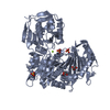
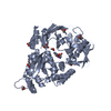
| Structure viewer | Molecule: MolmilJmol/JSmol |
|---|
- Downloads & links
-Download
| PDBx/mmCIF format | 3tpa.cif.gz | 228.9 KB | Display | PDBx/mmCIF format |
|---|---|---|---|---|
| PDB format | pdb3tpa.ent.gz | 181.9 KB | Display | PDB format |
| PDBx/mmJSON format | 3tpa.json.gz | Tree view | PDBx/mmJSON format | |
| Others |  Other downloads Other downloads |
-Validation report
| Arichive directory | https://data.pdbj.org/pub/pdb/validation_reports/tp/3tpaftp://data.pdbj.org/pub/pdb/validation_reports/tp/3tpa | HTTPS FTP |
|---|
-Related structure data
| Similar structure data |
|---|


-Links
 PDBj
PDBj- Assembly
| Deposited unit | 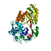
| ||||||||
|---|---|---|---|---|---|---|---|---|---|
| 1 |
| ||||||||
| Unit cell |
|
-Components
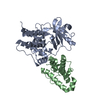
| #1: Protein | Mass: 58883.918 Da / Num. of mol.: 1 / Mutation: R290Q Y197N V453A T455L Source method: isolated from a genetically manipulated source Source: (gene. exp.) Haemophilus parasuis 29755 (bacteria) / Gene: HPS_10190 / Production host: Escherichia coli (E. coli) / References: UniProt: B0QSS4 |
|---|---|
| #2: Water | ChemComp-HOH / Water Mass: 18.015 Da / Num. of mol.: 637 / Source method: isolated from a natural source / Formula: H2O Mass: 18.015 Da / Num. of mol.: 637 / Source method: isolated from a natural source / Formula: H2O |
-Experimental details
-Experiment
| Experiment | Method: X-RAY DIFFRACTION / Number of used crystals: 1 |
|---|
- Sample preparation
| Crystal | Density Matthews: 2.13 Å3/Da / Density % sol: 42.26 % |
|---|---|
| Crystal grow | Temperature: 293 K / Method: vapor diffusion, sitting drop / pH: 7 Details: 0.1 M HEPES pH 7.0, 30% v/v jeffamine ED-2001, VAPOR DIFFUSION, SITTING DROP, temperature 293K |
-Data collection
| Diffraction | Mean temperature: 100 K |
|---|---|
| Diffraction source | Source: SYNCHROTRON / Site: SLS  / Beamline: X06DA / Wavelength: 1 Å / Beamline: X06DA / Wavelength: 1 Å |
| Detector | Type: MARMOSAIC 225 mm CCD / Detector: CCD |
| Radiation | Protocol: SINGLE WAVELENGTH / Monochromatic (M) / Laue (L): M / Scattering type: x-ray |
| Radiation wavelength | Wavelength: 1 Å / Relative weight: 1 |
| Reflection | Resolution: 2→50 Å / Num. all: 34932 / Num. obs: 34819 / % possible obs: 99.7 % |
| Reflection shell | Resolution: 2→2.05 Å / % possible obs: 99.8 % / Redundancy: 5.6 % / Mean I/σ(I) obs: 2.57 |
- Processing
| Software |
| |||||||||||||||||||||||||||||||||||||||||||||||||||||||||||||||||||||||||||||||||||||||||||
|---|---|---|---|---|---|---|---|---|---|---|---|---|---|---|---|---|---|---|---|---|---|---|---|---|---|---|---|---|---|---|---|---|---|---|---|---|---|---|---|---|---|---|---|---|---|---|---|---|---|---|---|---|---|---|---|---|---|---|---|---|---|---|---|---|---|---|---|---|---|---|---|---|---|---|---|---|---|---|---|---|---|---|---|---|---|---|---|---|---|---|---|---|
| Refinement | Method to determine structure: MOLECULAR REPLACEMENT Starting model: GBPA STRUCTURE FROM H. PARASUIS Resolution: 2.0001→40.639 Å / Occupancy max: 1 / Occupancy min: 0.39 / FOM work R set: 0.8691 / SU ML: 0.24 / σ(F): 2 / Phase error: 19.67 / Stereochemistry target values: ML
| |||||||||||||||||||||||||||||||||||||||||||||||||||||||||||||||||||||||||||||||||||||||||||
| Solvent computation | Shrinkage radii: 0.83 Å / VDW probe radii: 1.1 Å / Solvent model: FLAT BULK SOLVENT MODEL / Bsol: 43.202 Å2 / ksol: 0.3 e/Å3 | |||||||||||||||||||||||||||||||||||||||||||||||||||||||||||||||||||||||||||||||||||||||||||
| Displacement parameters | Biso max: 99.03 Å2 / Biso mean: 26.5604 Å2 / Biso min: 6.93 Å2
| |||||||||||||||||||||||||||||||||||||||||||||||||||||||||||||||||||||||||||||||||||||||||||
| Refine analyze | Luzzati coordinate error obs: 0.219 Å | |||||||||||||||||||||||||||||||||||||||||||||||||||||||||||||||||||||||||||||||||||||||||||
| Refinement step | Cycle: LAST / Resolution: 2.0001→40.639 Å
| |||||||||||||||||||||||||||||||||||||||||||||||||||||||||||||||||||||||||||||||||||||||||||
| Refine LS restraints |
| |||||||||||||||||||||||||||||||||||||||||||||||||||||||||||||||||||||||||||||||||||||||||||
| LS refinement shell | Refine-ID: X-RAY DIFFRACTION / Total num. of bins used: 12
| |||||||||||||||||||||||||||||||||||||||||||||||||||||||||||||||||||||||||||||||||||||||||||
| Refinement TLS params. | Method: refined / Refine-ID: X-RAY DIFFRACTION
| |||||||||||||||||||||||||||||||||||||||||||||||||||||||||||||||||||||||||||||||||||||||||||
| Refinement TLS group |
|
