 Movie
Movie Controller

[English] 日本語
 Yorodumi
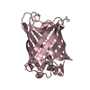
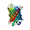
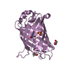
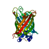
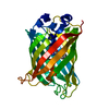
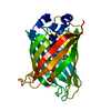
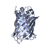
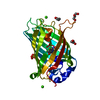
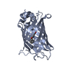
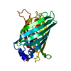
- PDB-5j2o: Crystal structure of the cyan fluorescence protein Cerulean S175G... -
+ Open data

- Basic information
| Entry | Database: PDB / ID: 5j2o | |||||||||
|---|---|---|---|---|---|---|---|---|---|---|
| Title | Crystal structure of the cyan fluorescence protein Cerulean S175G mutant | |||||||||
 Components Components | Green fluorescent protein | |||||||||
 Keywords Keywords | FLUORESCENT PROTEIN / Cerulean / Cyan Fluorescent Protein | |||||||||
| Function / homology |  Function and homology information Function and homology information | |||||||||
| Biological species |   Aequorea victoria (jellyfish) Aequorea victoria (jellyfish) | |||||||||
| Method | X-RAY DIFFRACTION / SYNCHROTRON / MOLECULAR REPLACEMENT / Resolution: 1.5 Å | |||||||||
 Authors Authors | Park, S.-W. / Kang, S. / Yoon, T.-S. | |||||||||
| Funding support |  Korea, Republic Of, 1items Korea, Republic Of, 1items
| |||||||||
 Citation Citation | Journal: Acta Crystallogr.,Sect.F / Year: 2016 Title: Crystal structure of the cyan fluorescent protein Cerulean-S175G Authors: Park, S.-W. / Kang, S. / Yoon, T.-S. | |||||||||
| History |
|
- Structure visualization
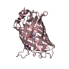
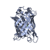
| Structure viewer | Molecule: MolmilJmol/JSmol |
|---|
- Downloads & links
-Download
| PDBx/mmCIF format | 5j2o.cif.gz | 102.6 KB | Display | PDBx/mmCIF format |
|---|---|---|---|---|
| PDB format | pdb5j2o.ent.gz | 78.2 KB | Display | PDB format |
| PDBx/mmJSON format | 5j2o.json.gz | Tree view | PDBx/mmJSON format | |
| Others |  Other downloads Other downloads |
-Validation report
| Arichive directory | https://data.pdbj.org/pub/pdb/validation_reports/j2/5j2oftp://data.pdbj.org/pub/pdb/validation_reports/j2/5j2o | HTTPS FTP |
|---|
-Related structure data
| Related structure data | 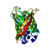 2ydzS S: Starting model for refinement |
|---|---|
| Similar structure data |


-Links
 PDBj
PDBj

- Assembly
| Deposited unit | 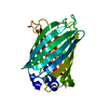
| ||||||||
|---|---|---|---|---|---|---|---|---|---|
| 1 |
| ||||||||
| Unit cell |
|
-Components
| #1: Protein | Mass: 27588.092 Da / Num. of mol.: 1 / Mutation: F65L/Y145A/N146I/H148D/M153T/V163A/S175G/H231L Source method: isolated from a genetically manipulated source Source: (gene. exp.) Aequorea victoria (jellyfish) / Gene: GFP / Plasmid: PET21A / Production host:  Escherichia coli (E. coli) / Strain (production host): BL21 CODONPLUS(DE3) / References: UniProt: P42212 Escherichia coli (E. coli) / Strain (production host): BL21 CODONPLUS(DE3) / References: UniProt: P42212 |
|---|---|
| #2: Water | ChemComp-HOH / Water Mass: 18.015 Da / Num. of mol.: 249 / Source method: isolated from a natural source / Formula: H2O Mass: 18.015 Da / Num. of mol.: 249 / Source method: isolated from a natural source / Formula: H2O |
| Sequence details | 1. S72A IS MUTAGENESIS ACCORDING TO DATABASE P42212 (GFP_AEQVI) 2. CHROMOPHORE RESULTING FROM ...1. S72A IS MUTAGENESI |
-Experimental details
-Experiment
| Experiment | Method: X-RAY DIFFRACTION / Number of used crystals: 1 |
|---|
- Sample preparation
| Crystal | Density Matthews: 2.04 Å3/Da / Density % sol: 39.81 % |
|---|---|
| Crystal grow | Temperature: 291 K / Method: vapor diffusion, sitting drop / pH: 7.5 / Details: 16% PEG 8000, 0.1M MGCL2, 0.1M HEPES PH 7.5 / PH range: 7.0-8.0 |
-Data collection
| Diffraction | Mean temperature: 100 K |
|---|---|
| Diffraction source | Source: SYNCHROTRON / Site: PAL/PLS / Beamline: 5C (4A) / Wavelength: 0.9796 Å |
| Detector | Type: ADSC QUANTUM 315r / Detector: CCD / Date: Jun 26, 2015 |
| Radiation | Monochromator: SI(111) / Protocol: SINGLE WAVELENGTH / Monochromatic (M) / Laue (L): M / Scattering type: x-ray |
| Radiation wavelength | Wavelength: 0.9796 Å / Relative weight: 1 |
| Reflection | Resolution: 1.5→35.053 Å / Num. obs: 36842 / % possible obs: 99.77 % / Redundancy: 7 % / Rsym value: 0.081 / Net I/av σ(I): 2.2 / Net I/σ(I): 24.3 |
| Reflection shell | Resolution: 1.5→1.53 Å / Redundancy: 0.249 % / Rmerge(I) obs: 0.249 / Mean I/σ(I) obs: 9 / % possible all: 100 |
- Processing
| Software |
| |||||||||||||||||||||||||||||||||||||||||||||||||||||||||||||||||||||||||||||||||||||||||||||||||||||||||
|---|---|---|---|---|---|---|---|---|---|---|---|---|---|---|---|---|---|---|---|---|---|---|---|---|---|---|---|---|---|---|---|---|---|---|---|---|---|---|---|---|---|---|---|---|---|---|---|---|---|---|---|---|---|---|---|---|---|---|---|---|---|---|---|---|---|---|---|---|---|---|---|---|---|---|---|---|---|---|---|---|---|---|---|---|---|---|---|---|---|---|---|---|---|---|---|---|---|---|---|---|---|---|---|---|---|---|
| Refinement | Method to determine structure: MOLECULAR REPLACEMENT Starting model: 2ydz Resolution: 1.5→35.053 Å / Cross valid method: FREE R-VALUE / ESU R Free: 0.1878 / Phase error: 96.33 / Details: add hydrogene
| |||||||||||||||||||||||||||||||||||||||||||||||||||||||||||||||||||||||||||||||||||||||||||||||||||||||||
| Solvent computation | Shrinkage radii: 0.9 Å / VDW probe radii: 1.11 Å / Bsol: 0.1411 Å2 / ksol: 0.9725 e/Å3 | |||||||||||||||||||||||||||||||||||||||||||||||||||||||||||||||||||||||||||||||||||||||||||||||||||||||||
| Displacement parameters | Biso max: 73.01 Å2 / Biso mean: 12.8524 Å2 / Biso min: 3.84 Å2 | |||||||||||||||||||||||||||||||||||||||||||||||||||||||||||||||||||||||||||||||||||||||||||||||||||||||||
| Refinement step | Cycle: LAST / Resolution: 1.5→35.053 Å
| |||||||||||||||||||||||||||||||||||||||||||||||||||||||||||||||||||||||||||||||||||||||||||||||||||||||||
| LS refinement shell | Refine-ID: X-RAY DIFFRACTION / Total num. of bins used: 14
|
