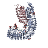
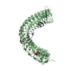
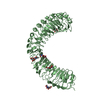
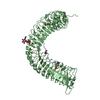
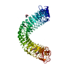
[English] 日本語
Yorodumi
- PDB-5ixo: Crystal structure of the Arabidopsis receptor kinase HAESA LRR ec... -

+
Open data


ID or keywords:

Loading...

-
Basic information

Entry
Database: PDB / ID: 5ixo
TitleCrystal structure of the Arabidopsis receptor kinase HAESA LRR ectdomain (apo form).
ComponentsReceptor-like protein kinase 5
KeywordsSIGNALING PROTEIN / membrane receptor kinase / peptide hormone receptor / signaling complex / plant development / organ shedding
Function / homology
Function and homology information


lateral root morphogenesis / leaf abscission / floral organ abscission / pectin catabolic process / transmembrane receptor protein tyrosine kinase activity / receptor protein-tyrosine kinase / regulation of gene expression / defense response to Gram-negative bacterium / protein autophosphorylation / non-specific serine/threonine protein kinase ...lateral root morphogenesis / leaf abscission / floral organ abscission / pectin catabolic process / transmembrane receptor protein tyrosine kinase activity / receptor protein-tyrosine kinase / regulation of gene expression / defense response to Gram-negative bacterium / protein autophosphorylation / non-specific serine/threonine protein kinase / protein kinase activity / protein serine kinase activity / protein serine/threonine kinase activity / ATP binding / plasma membrane
Similarity search - Function
Leucine-rich repeat-containing N-terminal, plant-type / Leucine rich repeat N-terminal domain / Leucine Rich Repeat / Leucine rich repeat / Leucine-rich repeat, typical subtype / Leucine-rich repeats, typical (most populated) subfamily / Leucine-rich repeat profile. / Leucine-rich repeat / Leucine-rich repeat domain superfamily / Serine/threonine-protein kinase, active site ...Leucine-rich repeat-containing N-terminal, plant-type / Leucine rich repeat N-terminal domain / Leucine Rich Repeat / Leucine rich repeat / Leucine-rich repeat, typical subtype / Leucine-rich repeats, typical (most populated) subfamily / Leucine-rich repeat profile. / Leucine-rich repeat / Leucine-rich repeat domain superfamily / Serine/threonine-protein kinase, active site / Serine/Threonine protein kinases active-site signature. / Protein kinase domain / Serine/Threonine protein kinases, catalytic domain / Protein kinase, ATP binding site / Protein kinases ATP-binding region signature. / Protein kinase domain profile. / Protein kinase domain / Protein kinase-like domain superfamily
Similarity search - Domain/homology
Receptor-like protein kinase 5
Similarity search - Component
Biological speciesArabidopsis thaliana (thale cress)
MethodX-RAY DIFFRACTION / SYNCHROTRON / SIRAS / Resolution: 1.74 Å
AuthorsSantiago, J. / Hothorn, M.
CitationJournal: Elife / Year: 2016
Title: Mechanistic insight into a peptide hormone signaling complex mediating floral organ abscission.
Authors: Santiago, J. / Brandt, B. / Wildhagen, M. / Hohmann, U. / Hothorn, L.A. / Butenko, M.A. / Hothorn, M.
History
DepositionMar 23, 2016Deposition site: RCSB / Processing site: PDBE
Revision 1.0Apr 20, 2016Provider: repository / Type: Initial release
Revision 2.0Jul 29, 2020Group: Atomic model / Data collection ...Atomic model / Data collection / Derived calculations / Structure summary
Category: atom_site / chem_comp ...atom_site / chem_comp / entity / pdbx_branch_scheme / pdbx_chem_comp_identifier / pdbx_entity_branch / pdbx_entity_branch_descriptor / pdbx_entity_branch_link / pdbx_entity_branch_list / pdbx_entity_nonpoly / pdbx_nonpoly_scheme / pdbx_struct_assembly_gen / struct_asym / struct_conn / struct_site / struct_site_gen
Item: _atom_site.B_iso_or_equiv / _atom_site.Cartn_x ..._atom_site.B_iso_or_equiv / _atom_site.Cartn_x / _atom_site.Cartn_y / _atom_site.Cartn_z / _atom_site.auth_asym_id / _atom_site.auth_seq_id / _atom_site.label_asym_id / _atom_site.label_entity_id / _chem_comp.name / _chem_comp.type / _pdbx_entity_nonpoly.entity_id / _pdbx_entity_nonpoly.name / _pdbx_struct_assembly_gen.asym_id_list / _struct_conn.conn_type_id / _struct_conn.id / _struct_conn.pdbx_dist_value / _struct_conn.pdbx_leaving_atom_flag / _struct_conn.pdbx_role / _struct_conn.ptnr1_auth_asym_id / _struct_conn.ptnr1_auth_comp_id / _struct_conn.ptnr1_auth_seq_id / _struct_conn.ptnr1_label_asym_id / _struct_conn.ptnr1_label_atom_id / _struct_conn.ptnr1_label_comp_id / _struct_conn.ptnr1_label_seq_id / _struct_conn.ptnr2_auth_asym_id / _struct_conn.ptnr2_auth_comp_id / _struct_conn.ptnr2_auth_seq_id / _struct_conn.ptnr2_label_asym_id / _struct_conn.ptnr2_label_atom_id / _struct_conn.ptnr2_label_comp_id
Description: Carbohydrate remediation / Provider: repository / Type: Remediation

-
Structure visualization

Structure viewerMolecule:
MolmilJmol/JSmol

Downloads & links

-
Assembly

Deposited unit
A: Receptor-like protein kinase 5
hetero molecules


Theoretical massNumber of molelcules
Total (without water)69,30110
Polymers67,2161
Non-polymers2,0859
Water1,40578
1


  • Idetical with deposited unit
  • defined by author&software
TypeNameSymmetry operationNumber
identity operation1_555x,y,z1
Buried area2550 Å2
ΔGint19 kcal/mol
Surface area25600 Å2
MethodPISA
Unit cell
Length a, b, c (Å)148.678, 148.678, 58.041
Angle α, β, γ (deg.)90.00, 90.00, 120.00
Int Tables number152
Space group name H-MP3121

-
Components

-
Protein , 1 types, 1 molecules A

#1: Protein Receptor-like protein kinase 5 / Protein HAESA


Mass: 67216.195 Da / Num. of mol.: 1 / Fragment: ectodomain, residues 20-620
Source method: isolated from a genetically manipulated source
Source: (gene. exp.) Arabidopsis thaliana (thale cress) / Tissue: abscission zone / Gene: RLK5, HAE, At4g28490, F21O9.180 / Organ: flower / Plasmid: pBAC3mod / Production host: Trichoplusia ni (cabbage looper) / Strain (production host): BTI38
References: UniProt: P47735, receptor protein-tyrosine kinase, non-specific serine/threonine protein kinase

-
Sugars , 2 types, 6 molecules

#2: Polysaccharide 2-acetamido-2-deoxy-beta-D-glucopyranose-(1-4)-2-acetamido-2-deoxy-beta-D-glucopyranose


Type: oligosaccharide / Mass: 424.401 Da / Num. of mol.: 3
Source method: isolated from a genetically manipulated source
DescriptorTypeProgram
DGlcpNAcb1-4DGlcpNAcb1-Glycam Condensed SequenceGMML 1.0
WURCS=2.0/1,2,1/[a2122h-1b_1-5_2*NCC/3=O]/1-1/a4-b1WURCSPDB2Glycan 1.1.0
[]{[(4+1)][b-D-GlcpNAc]{[(4+1)][b-D-GlcpNAc]{}}}LINUCSPDB-CARE
#3: Sugar ChemComp-NAG / 2-acetamido-2-deoxy-beta-D-glucopyranose / N-Acetylglucosamine


Type: D-saccharide, beta linking / Mass: 221.208 Da / Num. of mol.: 3
Source method: isolated from a genetically manipulated source
Formula: C8H15NO6
IdentifierTypeProgram
DGlcpNAcbCONDENSED IUPAC CARBOHYDRATE SYMBOLGMML 1.0
N-acetyl-b-D-glucopyranosamineCOMMON NAMEGMML 1.0
b-D-GlcpNAcIUPAC CARBOHYDRATE SYMBOLPDB-CARE 1.0
GlcNAcSNFG CARBOHYDRATE SYMBOLGMML 1.0

-
Non-polymers , 3 types, 81 molecules

#4: Chemical ChemComp-MG / MAGNESIUM ION


Mass: 24.305 Da / Num. of mol.: 1 / Source method: obtained synthetically / Formula: Mg
#5: Chemical ChemComp-EDO / 1,2-ETHANEDIOL / ETHYLENE GLYCOL / Ethylene glycol


Mass: 62.068 Da / Num. of mol.: 2 / Source method: obtained synthetically / Formula: C2H6O2
#6: Water ChemComp-HOH / water / Water


Mass: 18.015 Da / Num. of mol.: 78 / Source method: isolated from a natural source / Formula: H2O

-
Experimental details

-
Experiment

ExperimentMethod: X-RAY DIFFRACTION / Number of used crystals: 1

-
Sample preparation

CrystalDensity Matthews: 2.76 Å3/Da / Density % sol: 55.36 %
Crystal growTemperature: 298 K / Method: vapor diffusion, hanging drop / pH: 4 / Details: 22% PEG 3350, 0.2 MgCl2, 0.1 M citric acid pH 4.0

-
Data collection

DiffractionMean temperature: 100 K
Diffraction sourceSource: SYNCHROTRON / Site: SLS / Beamline: X06SA / Wavelength: 1.00001 Å
DetectorType: DECTRIS PILATUS3 6M / Detector: PIXEL / Date: Mar 23, 2014
RadiationProtocol: SINGLE WAVELENGTH / Monochromatic (M) / Laue (L): M / Scattering type: x-ray
Radiation wavelengthWavelength: 1.00001 Å / Relative weight: 1
ReflectionResolution: 1.74→128.76 Å / Num. obs: 74928 / % possible obs: 99.6 % / Observed criterion σ(F): -3 / Redundancy: 14.4 % / Biso Wilson estimate: 81.1 Å2 / CC1/2: 1 / Rsym value: 0.048 / Net I/σ(I): 18.7
Reflection shellResolution: 1.74→1.85 Å / Redundancy: 14 % / Mean I/σ(I) obs: 1.8 / % possible all: 97.4

-
Processing

Software
NameVersionClassification
REFMAC5.8.0135refinement
XDSdata reduction
XSCALEdata scaling
SHARPphasing
RefinementMethod to determine structure: SIRAS / Resolution: 1.74→128.76 Å / Cor.coef. Fo:Fc: 0.976 / Cor.coef. Fo:Fc free: 0.964 / SU B: 7.857 / SU ML: 0.111 / Cross valid method: THROUGHOUT / ESU R: 0.096 / ESU R Free: 0.102 / Details: HYDROGENS HAVE BEEN ADDED IN THE RIDING POSITIONS
RfactorNum. reflection% reflectionSelection details
Rfree0.22172 3713 5 %RANDOM
Rwork0.1802 ---
obs0.18227 71213 99.63 %-
Solvent computationIon probe radii: 0.8 Å / Shrinkage radii: 0.8 Å / VDW probe radii: 1.2 Å
Displacement parametersBiso mean: 80.252 Å2
Baniso -1Baniso -2Baniso -3
1-1.47 Å20.74 Å20 Å2
2--1.47 Å2-0 Å2
3----4.78 Å2
Refinement stepCycle: 1 / Resolution: 1.74→128.76 Å
ProteinNucleic acidLigandSolventTotal
Num. atoms4508 0 135 78 4721
Refine LS restraints
Refine-IDTypeDev idealDev ideal targetNumber
X-RAY DIFFRACTIONr_bond_refined_d0.0250.0194762
X-RAY DIFFRACTIONr_bond_other_d0.0020.024536
X-RAY DIFFRACTIONr_angle_refined_deg2.2592.0226488
X-RAY DIFFRACTIONr_angle_other_deg1.173310497
X-RAY DIFFRACTIONr_dihedral_angle_1_deg6.5185603
X-RAY DIFFRACTIONr_dihedral_angle_2_deg43.56326.022186
X-RAY DIFFRACTIONr_dihedral_angle_3_deg12.99915790
X-RAY DIFFRACTIONr_dihedral_angle_4_deg18.4511514
X-RAY DIFFRACTIONr_chiral_restr0.1470.2775
X-RAY DIFFRACTIONr_gen_planes_refined0.0120.0215354
X-RAY DIFFRACTIONr_gen_planes_other0.0020.02990
X-RAY DIFFRACTIONr_nbd_refined
X-RAY DIFFRACTIONr_nbd_other
X-RAY DIFFRACTIONr_nbtor_refined
X-RAY DIFFRACTIONr_nbtor_other
X-RAY DIFFRACTIONr_xyhbond_nbd_refined
X-RAY DIFFRACTIONr_xyhbond_nbd_other
X-RAY DIFFRACTIONr_metal_ion_refined
X-RAY DIFFRACTIONr_metal_ion_other
X-RAY DIFFRACTIONr_symmetry_vdw_refined
X-RAY DIFFRACTIONr_symmetry_vdw_other
X-RAY DIFFRACTIONr_symmetry_hbond_refined
X-RAY DIFFRACTIONr_symmetry_hbond_other
X-RAY DIFFRACTIONr_symmetry_metal_ion_refined
X-RAY DIFFRACTIONr_symmetry_metal_ion_other
X-RAY DIFFRACTIONr_mcbond_it3.7065.6232391
X-RAY DIFFRACTIONr_mcbond_other3.6965.6232390
X-RAY DIFFRACTIONr_mcangle_it4.2278.4082989
X-RAY DIFFRACTIONr_mcangle_other4.238.4082990
X-RAY DIFFRACTIONr_scbond_it5.766.2952368
X-RAY DIFFRACTIONr_scbond_other5.766.2952368
X-RAY DIFFRACTIONr_scangle_it
X-RAY DIFFRACTIONr_scangle_other7.6099.2443495
X-RAY DIFFRACTIONr_long_range_B_refined8.35555.13720122
X-RAY DIFFRACTIONr_long_range_B_other8.35655.13220116
X-RAY DIFFRACTIONr_rigid_bond_restr
X-RAY DIFFRACTIONr_sphericity_free
X-RAY DIFFRACTIONr_sphericity_bonded
LS refinement shellResolution: 1.743→1.788 Å / Total num. of bins used: 20
RfactorNum. reflection% reflection
Rfree0.543 321 -
Rwork0.514 4952 -
obs--95.15 %
Refinement TLS params.

Method: refined / Refine-ID: X-RAY DIFFRACTION

IDL112)L122)L132)L222)L232)L332)S11 (Å °)S12 (Å °)S13 (Å °)S21 (Å °)S22 (Å °)S23 (Å °)S31 (Å °)S32 (Å °)S33 (Å °)T112)T122)T132)T222)T232)T332)Origin x (Å)Origin y (Å)Origin z (Å)
15.9250.2556-0.28552.8558-0.05916.1967-0.0588-0.3730.5647-0.0552-0.09640.4678-0.6708-0.27460.15520.10550.0725-0.07350.12690.0130.4669-13.65884.812-14.999
21.2349-0.97930.43632.3734-0.69820.8730.04020.11570.11-0.1947-0.0786-0.01590.01390.02560.03840.02070.00290.01140.01130.00540.36288.73365.946-11.129
31.69110.6734-0.08892.10790.13032.28540.1595-0.28460.05610.2162-0.1990.03980.0023-0.0930.03950.029-0.03480.01710.0572-0.01230.35179.72147.70220.57
43.68452.80250.84393.70350.16491.16060.4631-0.6920.12560.1636-0.51620.31140.1159-0.30050.05310.1269-0.15320.04760.3339-0.02580.3759-20.31426.68921.611
Refinement TLS group
IDRefine-IDRefine TLS-IDAuth asym-IDAuth seq-ID
1X-RAY DIFFRACTION1A20 - 55
2X-RAY DIFFRACTION2A56 - 275
3X-RAY DIFFRACTION3A276 - 423
4X-RAY DIFFRACTION4A424 - 615

+
About Yorodumi

-
News

-
Feb 9, 2022. New format data for meta-information of EMDB entries

New format data for meta-information of EMDB entries

  • Version 3 of the EMDB header file is now the official format.
  • The previous official version 1.9 will be removed from the archive.

Related info.:EMDB header

External links:wwPDB to switch to version 3 of the EMDB data model

-
Aug 12, 2020. Covid-19 info

Covid-19 info

URL: https://pdbj.org/emnavi/covid19.php

New page: Covid-19 featured information page in EM Navigator.

Related info.:Covid-19 info / Mar 5, 2020. Novel coronavirus structure data

+
Mar 5, 2020. Novel coronavirus structure data

Novel coronavirus structure data

Related info.:Yorodumi Speices / Aug 12, 2020. Covid-19 info

External links:COVID-19 featured content - PDBj / Molecule of the Month (242):Coronavirus Proteases

+
Jan 31, 2019. EMDB accession codes are about to change! (news from PDBe EMDB page)

EMDB accession codes are about to change! (news from PDBe EMDB page)

  • The allocation of 4 digits for EMDB accession codes will soon come to an end. Whilst these codes will remain in use, new EMDB accession codes will include an additional digit and will expand incrementally as the available range of codes is exhausted. The current 4-digit format prefixed with “EMD-” (i.e. EMD-XXXX) will advance to a 5-digit format (i.e. EMD-XXXXX), and so on. It is currently estimated that the 4-digit codes will be depleted around Spring 2019, at which point the 5-digit format will come into force.
  • The EM Navigator/Yorodumi systems omit the EMD- prefix.

Related info.:Q: What is EMD? / ID/Accession-code notation in Yorodumi/EM Navigator

External links:EMDB Accession Codes are Changing Soon! / Contact to PDBj

+
Jul 12, 2017. Major update of PDB

Major update of PDB

  • wwPDB released updated PDB data conforming to the new PDBx/mmCIF dictionary.
  • This is a major update changing the version number from 4 to 5, and with Remediation, in which all the entries are updated.
  • In this update, many items about electron microscopy experimental information are reorganized (e.g. em_software).
  • Now, EM Navigator and Yorodumi are based on the updated data.

External links:wwPDB Remediation / Enriched Model Files Conforming to OneDep Data Standards Now Available in the PDB FTP Archive

-
Yorodumi

Thousand views of thousand structures

  • Yorodumi is a browser for structure data from EMDB, PDB, SASBDB, etc.
  • This page is also the successor to EM Navigator detail page, and also detail information page/front-end page for Omokage search.
  • The word "yorodu" (or yorozu) is an old Japanese word meaning "ten thousand". "mi" (miru) is to see.

Related info.:EMDB / PDB / SASBDB / Comparison of 3 databanks / Yorodumi Search / Aug 31, 2016. New EM Navigator & Yorodumi / Yorodumi Papers / Jmol/JSmol / Function and homology information / Changes in new EM Navigator and Yorodumi

Read more