 Movie
Movie Controller

[English] 日本語
 Yorodumi
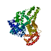
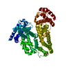
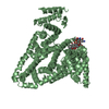
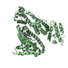
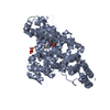
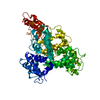
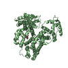
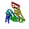
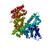
- PDB-5iiu: Crystal structure of Equine Serum Albumin in the presence of 10 m... -
+ Open data

- Basic information
| Entry | Database: PDB / ID: 5iiu | ||||||||||||
|---|---|---|---|---|---|---|---|---|---|---|---|---|---|
| Title | Crystal structure of Equine Serum Albumin in the presence of 10 mM zinc at pH 6.9 | ||||||||||||
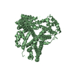
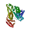
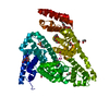
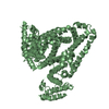
 Components Components | Serum albumin | ||||||||||||
 Keywords Keywords | TRANSPORT PROTEIN / Structural Genomics / PSI-Biology / New York Structural Genomics Research Consortium / NYSGRC | ||||||||||||
| Function / homology |  Function and homology information Function and homology informationcellular response to calcium ion starvation / enterobactin binding / negative regulation of mitochondrial depolarization / toxic substance binding / cellular response to starvation / fatty acid binding / pyridoxal phosphate binding / protein-containing complex / DNA binding / extracellular space ...cellular response to calcium ion starvation / enterobactin binding / negative regulation of mitochondrial depolarization / toxic substance binding / cellular response to starvation / fatty acid binding / pyridoxal phosphate binding / protein-containing complex / DNA binding / extracellular space / metal ion binding / cytoplasmSimilarity search - Function | ||||||||||||
| Biological species |  Equus caballus (horse) Equus caballus (horse) | ||||||||||||
| Method | X-RAY DIFFRACTION / SYNCHROTRON / MOLECULAR REPLACEMENT / Resolution: 2.3 Å | ||||||||||||
 Authors Authors | Handing, K.B. / Shabalin, I.G. / Cooper, D.R. / Cymborowski, M.T. / Almo, S.C. / Minor, W. / New York Structural Genomics Research Consortium (NYSGRC) | ||||||||||||
| Funding support |  United States, 3items United States, 3items
| ||||||||||||
 Citation Citation | Journal: Chem Sci / Year: 2016 Title: Circulatory zinc transport is controlled by distinct interdomain sites on mammalian albumins. Authors: Handing, K.B. / Shabalin, I.G. / Kassaar, O. / Khazaipoul, S. / Blindauer, C.A. / Stewart, A.J. / Chruszcz, M. / Minor, W. | ||||||||||||
| History |
|
- Structure visualization
| Structure viewer | Molecule: MolmilJmol/JSmol |
|---|
- Downloads & links
-Download
| PDBx/mmCIF format | 5iiu.cif.gz | 247.2 KB | Display | PDBx/mmCIF format |
|---|---|---|---|---|
| PDB format | pdb5iiu.ent.gz | 198.5 KB | Display | PDB format |
| PDBx/mmJSON format | 5iiu.json.gz | Tree view | PDBx/mmJSON format | |
| Others |  Other downloads Other downloads |
-Validation report
| Arichive directory | https://data.pdbj.org/pub/pdb/validation_reports/ii/5iiuftp://data.pdbj.org/pub/pdb/validation_reports/ii/5iiu | HTTPS FTP |
|---|
-Related structure data
| Related structure data |  5iihSC 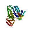 5iixC 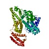 5ij5C  5ijeC 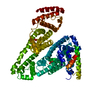 5ijfC S: Starting model for refinement C: citing same article ( |
|---|---|
| Similar structure data | |
| Other databases |


-Links
 PDBj
PDBj


- Assembly
| Deposited unit | 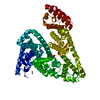
| ||||||||
|---|---|---|---|---|---|---|---|---|---|
| 1 |
| ||||||||
| Unit cell |
|
-Components
| #1: Protein | Mass: 65768.086 Da / Num. of mol.: 1 / Fragment: residues 25-607 / Source method: isolated from a natural source / Source: (natural) Equus caballus (horse) / References: UniProt: P35747 | ||||||
|---|---|---|---|---|---|---|---|
| #2: Chemical | ChemComp-ZN /   Mass: 65.409 Da / Num. of mol.: 10 / Source method: obtained synthetically / Formula: Zn Mass: 65.409 Da / Num. of mol.: 10 / Source method: obtained synthetically / Formula: Zn#3: Chemical | ChemComp-SO4 / | Sulfate  Mass: 96.063 Da / Num. of mol.: 1 / Source method: obtained synthetically / Formula: SO4 Mass: 96.063 Da / Num. of mol.: 1 / Source method: obtained synthetically / Formula: SO4#4: Water | ChemComp-HOH / | Water Mass: 18.015 Da / Num. of mol.: 193 / Source method: isolated from a natural source / Formula: H2O Mass: 18.015 Da / Num. of mol.: 193 / Source method: isolated from a natural source / Formula: H2OSequence details | In contrast with the previously deposited in PDB structures of Equus caballus SA (PDB IDs: 3V08, ...In contrast with the previously deposited in PDB structures of Equus caballus SA (PDB IDs: 3V08, 4J2V, 4OT2, 4F5U, and 4F5T), a single point mutation, R561A, is observed. The long arginine side chain cannot be modeled in this position due to steric clashes with the nearby disulfide bond connecting Cys567 and Cys558 and a symmetry-related copy of the molecule. Moreover, there is no 2mFo-DFc omit map supporting placement of the side chain. Protein was purified from natural source, therefore there may be naturally occurring mutation. According to the NCBI database, this mutation is characteristic for Equus ferus przewalskii, a rare subspecies of wild horse from central Asia (accession code: XP_008524663.1). However it is possible that there is an error in the Equus caballus SA sequence, or the observed mutation naturally occurs in that species. | |
-Experimental details
-Experiment
| Experiment | Method: X-RAY DIFFRACTION / Number of used crystals: 1 |
|---|
- Sample preparation
| Crystal | Density Matthews: 2.73 Å3/Da / Density % sol: 55.02 % |
|---|---|
| Crystal grow | Temperature: 289 K / Method: vapor diffusion, hanging drop / pH: 6.9 Details: 1 ul of 30 mg/ml protein in 10 mM Tris pH 7.5 and 150 mM NaCl buffer was mixed with 1 ul of the well condition (0.2 M Li2SO4, 0.1 M Tris:HCl, 2.0 M (NH4)2SO4, 5 mM ZnCl2, final pH 6.9) and ...Details: 1 ul of 30 mg/ml protein in 10 mM Tris pH 7.5 and 150 mM NaCl buffer was mixed with 1 ul of the well condition (0.2 M Li2SO4, 0.1 M Tris:HCl, 2.0 M (NH4)2SO4, 5 mM ZnCl2, final pH 6.9) and equilibrated against well solution in 15 Well Crystallization Plate (Qiagen). Crystals were soaked with 50 mM ZnCl2 in 100 mM Tris, final pH 6.9, to final concentration of 10 mM |
-Data collection
| Diffraction | Mean temperature: 100 K | |||||||||||||||||||||||||||||||||||||||||||||||||||||||||||||||||||||||||||||||||||||||||||||||||||||||||
|---|---|---|---|---|---|---|---|---|---|---|---|---|---|---|---|---|---|---|---|---|---|---|---|---|---|---|---|---|---|---|---|---|---|---|---|---|---|---|---|---|---|---|---|---|---|---|---|---|---|---|---|---|---|---|---|---|---|---|---|---|---|---|---|---|---|---|---|---|---|---|---|---|---|---|---|---|---|---|---|---|---|---|---|---|---|---|---|---|---|---|---|---|---|---|---|---|---|---|---|---|---|---|---|---|---|---|
| Diffraction source | Source: SYNCHROTRON / Site: APS / Beamline: 21-ID-G / Wavelength: 0.979 Å | |||||||||||||||||||||||||||||||||||||||||||||||||||||||||||||||||||||||||||||||||||||||||||||||||||||||||
| Detector | Type: MARMOSAIC 300 mm CCD / Detector: CCD / Date: Dec 4, 2014 / Details: Beryllium Lenses | |||||||||||||||||||||||||||||||||||||||||||||||||||||||||||||||||||||||||||||||||||||||||||||||||||||||||
| Radiation | Monochromator: Diamond [111] / Protocol: SINGLE WAVELENGTH / Monochromatic (M) / Laue (L): M / Scattering type: x-ray | |||||||||||||||||||||||||||||||||||||||||||||||||||||||||||||||||||||||||||||||||||||||||||||||||||||||||
| Radiation wavelength | Wavelength: 0.979 Å / Relative weight: 1 | |||||||||||||||||||||||||||||||||||||||||||||||||||||||||||||||||||||||||||||||||||||||||||||||||||||||||
| Reflection | Resolution: 2.3→80.01 Å / Num. obs: 31099 / % possible obs: 99.3 % / Observed criterion σ(I): -3 / Redundancy: 9.7 % / Biso Wilson estimate: 49.5 Å2 / Rmerge(I) obs: 0.078 / Rsym value: 0.078 / Net I/av σ(I): 29.091 / Net I/σ(I): 7.6 | |||||||||||||||||||||||||||||||||||||||||||||||||||||||||||||||||||||||||||||||||||||||||||||||||||||||||
| Reflection shell |
|
- Processing
| Software |
| |||||||||||||||||||||||||||||||||||||||||||||||||||||||||||||||||||||||||||||||||||||||||||||||||||||||||||||||||||||||||||||||||||||||||||||||||||||||||||||||||||||||||||||||
|---|---|---|---|---|---|---|---|---|---|---|---|---|---|---|---|---|---|---|---|---|---|---|---|---|---|---|---|---|---|---|---|---|---|---|---|---|---|---|---|---|---|---|---|---|---|---|---|---|---|---|---|---|---|---|---|---|---|---|---|---|---|---|---|---|---|---|---|---|---|---|---|---|---|---|---|---|---|---|---|---|---|---|---|---|---|---|---|---|---|---|---|---|---|---|---|---|---|---|---|---|---|---|---|---|---|---|---|---|---|---|---|---|---|---|---|---|---|---|---|---|---|---|---|---|---|---|---|---|---|---|---|---|---|---|---|---|---|---|---|---|---|---|---|---|---|---|---|---|---|---|---|---|---|---|---|---|---|---|---|---|---|---|---|---|---|---|---|---|---|---|---|---|---|---|---|---|
| Refinement | Method to determine structure: MOLECULAR REPLACEMENT Starting model: 5IIH Resolution: 2.3→80.01 Å / Cor.coef. Fo:Fc: 0.965 / Cor.coef. Fo:Fc free: 0.935 / SU B: 16.572 / SU ML: 0.2 / SU R Cruickshank DPI: 0.2776 / Cross valid method: THROUGHOUT / σ(F): 0 / ESU R: 0.278 / ESU R Free: 0.224 Details: U VALUES : WITH TLS ADDED HYDROGENS HAVE BEEN ADDED IN THE RIDING POSITIONS
| |||||||||||||||||||||||||||||||||||||||||||||||||||||||||||||||||||||||||||||||||||||||||||||||||||||||||||||||||||||||||||||||||||||||||||||||||||||||||||||||||||||||||||||||
| Solvent computation | Ion probe radii: 0.8 Å / Shrinkage radii: 0.8 Å / VDW probe radii: 1.2 Å | |||||||||||||||||||||||||||||||||||||||||||||||||||||||||||||||||||||||||||||||||||||||||||||||||||||||||||||||||||||||||||||||||||||||||||||||||||||||||||||||||||||||||||||||
| Displacement parameters | Biso max: 153.04 Å2 / Biso mean: 61.773 Å2 / Biso min: 32.33 Å2
| |||||||||||||||||||||||||||||||||||||||||||||||||||||||||||||||||||||||||||||||||||||||||||||||||||||||||||||||||||||||||||||||||||||||||||||||||||||||||||||||||||||||||||||||
| Refinement step | Cycle: final / Resolution: 2.3→80.01 Å
| |||||||||||||||||||||||||||||||||||||||||||||||||||||||||||||||||||||||||||||||||||||||||||||||||||||||||||||||||||||||||||||||||||||||||||||||||||||||||||||||||||||||||||||||
| Refine LS restraints |
| |||||||||||||||||||||||||||||||||||||||||||||||||||||||||||||||||||||||||||||||||||||||||||||||||||||||||||||||||||||||||||||||||||||||||||||||||||||||||||||||||||||||||||||||
| LS refinement shell | Resolution: 2.301→2.361 Å / Total num. of bins used: 20
| |||||||||||||||||||||||||||||||||||||||||||||||||||||||||||||||||||||||||||||||||||||||||||||||||||||||||||||||||||||||||||||||||||||||||||||||||||||||||||||||||||||||||||||||
| Refinement TLS params. | Method: refined / Refine-ID: X-RAY DIFFRACTION
| |||||||||||||||||||||||||||||||||||||||||||||||||||||||||||||||||||||||||||||||||||||||||||||||||||||||||||||||||||||||||||||||||||||||||||||||||||||||||||||||||||||||||||||||
| Refinement TLS group |
|
