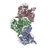
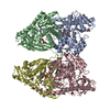
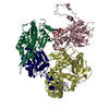
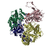
Quantitative interaction mapping reveals an extended ubiquitin regulatory domain in ASPL that disrupts functional p97 hexamers and induces cell death
Components
Tether containing UBX domain for GLUT4
Transitional endoplasmic reticulum ATPase
Keywords
SIGNALING PROTEIN / ASPL / p97 / disassembly / eUBX
Function / homology
Function and homology information
positive regulation of protein modification process / regulation of glucose import / positive regulation of Lys63-specific deubiquitinase activity / flavin adenine dinucleotide catabolic process / positive regulation of oxidative phosphorylation / VCP-NSFL1C complex / vesicle membrane / endosome to lysosome transport via multivesicular body sorting pathway / protein-DNA covalent cross-linking repair / endoplasmic reticulum stress-induced pre-emptive quality control ...positive regulation of protein modification process / regulation of glucose import / positive regulation of Lys63-specific deubiquitinase activity / flavin adenine dinucleotide catabolic process / positive regulation of oxidative phosphorylation / VCP-NSFL1C complex / vesicle membrane / endosome to lysosome transport via multivesicular body sorting pathway / protein-DNA covalent cross-linking repair / endoplasmic reticulum stress-induced pre-emptive quality control / cellular response to arsenite ion / Derlin-1 retrotranslocation complex / BAT3 complex binding / positive regulation of protein K63-linked deubiquitination / deubiquitinase activator activity / mitotic spindle disassembly / VCP-NPL4-UFD1 AAA ATPase complex / aggresome assembly / regulation of protein localization to chromatin / vesicle-fusing ATPase / NADH metabolic process / cellular response to misfolded protein / stress granule disassembly / K48-linked polyubiquitin modification-dependent protein binding / positive regulation of mitochondrial membrane potential / negative regulation of protein localization to chromatin / ubiquitin-modified protein reader activity / retrograde protein transport, ER to cytosol / regulation of aerobic respiration / ATPase complex / regulation of synapse organization / ubiquitin-specific protease binding / positive regulation of ATP biosynthetic process / ERAD pathway / ubiquitin-like protein ligase binding / MHC class I protein binding / autophagosome maturation / RHOH GTPase cycle / polyubiquitin modification-dependent protein binding / HSF1 activation / endomembrane system / translesion synthesis / Protein methylation / endoplasmic reticulum to Golgi vesicle-mediated transport / interstrand cross-link repair / negative regulation of smoothened signaling pathway / ATP metabolic process / Attachment and Entry / endoplasmic reticulum unfolded protein response / endoplasmic reticulum-Golgi intermediate compartment membrane / viral genome replication / lipid droplet / proteasome complex / proteasomal protein catabolic process / Josephin domain DUBs / N-glycan trimming in the ER and Calnexin/Calreticulin cycle / Translocation of SLC2A4 (GLUT4) to the plasma membrane / Hh mutants are degraded by ERAD / Defective CFTR causes cystic fibrosis / Hedgehog ligand biogenesis / macroautophagy / intracellular protein transport / Translesion Synthesis by POLH / positive regulation of protein-containing complex assembly / ABC-family proteins mediated transport / establishment of protein localization / ADP binding / cytoplasmic side of plasma membrane / autophagy / Aggrephagy / cytoplasmic stress granule / positive regulation of non-canonical NF-kappaB signal transduction / positive regulation of protein catabolic process / activation of cysteine-type endopeptidase activity involved in apoptotic process / KEAP1-NFE2L2 pathway / azurophil granule lumen / double-strand break repair / positive regulation of canonical Wnt signaling pathway / Ovarian tumor domain proteases / positive regulation of proteasomal ubiquitin-dependent protein catabolic process / E3 ubiquitin ligases ubiquitinate target proteins / glucose homeostasis / site of double-strand break / Neddylation / cellular response to heat / ubiquitin-dependent protein catabolic process / regulation of apoptotic process / protein phosphatase binding / proteasome-mediated ubiquitin-dependent protein catabolic process / secretory granule lumen / ficolin-1-rich granule lumen / Attachment and Entry / protein ubiquitination / protein domain specific binding / intracellular membrane-bounded organelle / DNA repair / glutamatergic synapse / lipid binding / DNA damage response / ubiquitin protein ligase binding Similarity search - Function
TethercontainingUBXdomainforGLUT4 / Alveolar soft part sarcoma chromosomal region candidate gene 1 protein / Alveolar soft part sarcoma ...Alveolar soft part sarcoma chromosomal region candidate gene 1 protein / Alveolar soft part sarcoma locus / Renal papillary cell carcinoma protein 17 / UBX domain-containing protein 9
Mass: 26451.355 Da / Num. of mol.: 2 / Fragment: UNP residues 317-553 Source method: isolated from a genetically manipulated source Source: (gene. exp.) Homo sapiens (human) / Gene: ASPSCR1, ASPL, RCC17, TUG, UBXD9, UBXN9 / Plasmid: pQLinkG / Production host: Escherichia coli BL21(DE3) (bacteria) / Variant (production host): Rosetta2 / References: UniProt: Q9BZE9
#2: Protein
TransitionalendoplasmicreticulumATPase / TER ATPase / 15S Mg(2+)-ATPase p97 subunit / Valosin-containing protein / VCP
Mass: 53508.168 Da / Num. of mol.: 2 Source method: isolated from a genetically manipulated source Source: (gene. exp.) Homo sapiens (human) / Gene: VCP / Plasmid: pQLinkG / Production host: Escherichia coli BL21(DE3) (bacteria) / Variant (production host): Rosetta 2 / References: UniProt: P55072, vesicle-fusing ATPase
Resolution: 2.46→33.77 Å / Cor.coef. Fo:Fc: 0.944 / Cor.coef. Fo:Fc free: 0.914 / SU B: 9.238 / SU ML: 0.205 / Cross valid method: THROUGHOUT / ESU R: 0.429 / ESU R Free: 0.272 / Stereochemistry target values: MAXIMUM LIKELIHOOD / Details: HYDROGENS HAVE BEEN USED IF PRESENT IN THE INPUT
Rfactor
Num. reflection
% reflection
Selection details
Rfree
0.25061
2930
5 %
RANDOM
Rwork
0.20101
-
-
-
obs
0.20352
55653
98.05 %
-
Solvent computation
Ion probe radii: 0.8 Å / Shrinkage radii: 0.8 Å / VDW probe radii: 1.2 Å / Solvent model: MASK
 Movie
Movie Controller

 Yorodumi
 Open data
 Basic information
 Components
 Keywords
 SIGNALING PROTEIN /
  Function and homology information
 Authors
 Citation
 Structure visualization
 Downloads & links
 Other downloads
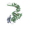


 PDBj
PDBj


 Assembly


 Mass: 427.201 Da / Num. of mol.: 2 / Source method: obtained synthetically / Formula: C10H15N5O10P2 / Comment: ADP, energy-carrying molecule*YM
 Mass: 24.305 Da / Num. of mol.: 2 / Source method: obtained synthetically / Formula: Mg
 Mass: 96.063 Da / Num. of mol.: 1 / Source method: obtained synthetically / Formula: SO4
 Mass: 62.068 Da / Num. of mol.: 3 / Source method: obtained synthetically / Formula: C2H6O2
 Mass: 92.094 Da / Num. of mol.: 1 / Source method: obtained synthetically / Formula: C3H8O3
 Sample preparation
 / Beamline: 14.1 / Wavelength: 0.918 Å
 Processing