 Movie
Movie Controller

[English] 日本語
 Yorodumi
- PDB-3oe6: Crystal structure of the CXCR4 chemokine receptor in complex with... -
+ Open data

- Basic information
| Entry | Database: PDB / ID: 3oe6 | ||||||
|---|---|---|---|---|---|---|---|
| Title | Crystal structure of the CXCR4 chemokine receptor in complex with a small molecule antagonist IT1t in I222 spacegroup | ||||||
 Components Components | C-X-C chemokine receptor type 4, Lysozyme Chimera | ||||||
 Keywords Keywords |  SIGNALING PROTEIN / HYDROLASE / Structural Genomics / PSI-2 / Protein Structure Initiative / Accelerated Technologies Center for Gene to 3D Structure / ATCG3D / 7TM / G protein-coupled receptor / GPCR / Signal transduction / Cancer / HIV-1 co-receptor / Chemokine / CXCL12 / SDF1 / isothiourea / IT1t / Chimera / T4L Fusion / Membrane protein / Transmembrane / SINGNALING PROTEIN / PSI-Biology / GPCR Network SIGNALING PROTEIN / HYDROLASE / Structural Genomics / PSI-2 / Protein Structure Initiative / Accelerated Technologies Center for Gene to 3D Structure / ATCG3D / 7TM / G protein-coupled receptor / GPCR / Signal transduction / Cancer / HIV-1 co-receptor / Chemokine / CXCL12 / SDF1 / isothiourea / IT1t / Chimera / T4L Fusion / Membrane protein / Transmembrane / SINGNALING PROTEIN / PSI-Biology / GPCR Network | ||||||
| Function / homology |  Function and homology information Function and homology informationC-X-C motif chemokine 12 receptor activity / regulation of viral process / positive regulation of vascular wound healing / positive regulation of macrophage migration inhibitory factor signaling pathway / positive regulation of mesenchymal stem cell migration / neuron recognition / response to ultrasound / response to tacrolimus / telencephalon cell migration / C-X-C chemokine receptor activity ...C-X-C motif chemokine 12 receptor activity / regulation of viral process / positive regulation of vascular wound healing / positive regulation of macrophage migration inhibitory factor signaling pathway / positive regulation of mesenchymal stem cell migration / neuron recognition / response to ultrasound / response to tacrolimus / telencephalon cell migration / C-X-C chemokine receptor activity / Specification of primordial germ cells / CXCL12-activated CXCR4 signaling pathway / myosin light chain binding / myelin maintenance / positive regulation of vasculature development / regulation of programmed cell death / C-C chemokine receptor activity / endothelial tube morphogenesis / endothelial cell differentiation / C-C chemokine binding / Signaling by ROBO receptors / regulation of chemotaxis / cellular response to organonitrogen compound / positive regulation of chemotaxis / Formation of definitive endoderm / positive regulation of dendrite extension / anchoring junction / Chemokine receptors bind chemokines / dendritic cell chemotaxis / positive regulation of oligodendrocyte differentiation / epithelial cell development / cell leading edge / cellular response to cytokine stimulus / small molecule binding / detection of temperature stimulus involved in sensory perception of pain / regulation of calcium ion transport / Binding and entry of HIV virion / coreceptor activity / detection of mechanical stimulus involved in sensory perception of pain / regulation of cell adhesion / cardiac muscle contraction / viral release from host cell by cytolysis / neurogenesis / cell chemotaxis / peptidoglycan catabolic process / response to activity / ubiquitin binding / G protein-coupled receptor activity / calcium-mediated signaling / brain development / neuron migration / response to virus / cell wall macromolecule catabolic process / cellular response to xenobiotic stimulus / late endosome / virus receptor activity / lysozyme / lysozyme activity / positive regulation of cold-induced thermogenesis / actin binding / positive regulation of cytosolic calcium ion concentration / G alpha (i) signalling events / cytoplasmic vesicle / host cell cytoplasm / lysosome / response to hypoxia / early endosome / positive regulation of cell migration / defense response to bacterium / immune response / inflammatory response / G protein-coupled receptor signaling pathway / external side of plasma membrane / apoptotic process / ubiquitin protein ligase binding / cell surface / protein-containing complex / extracellular exosome / plasma membrane / cytoplasmSimilarity search - Function | ||||||
| Biological species |  Homo Sapiens (human) Homo Sapiens (human) Enterobacteria phage T4 (virus) Enterobacteria phage T4 (virus) | ||||||
| Method | X-RAY DIFFRACTION / SYNCHROTRON / MOLECULAR REPLACEMENT / Resolution: 3.2 Å | ||||||
 Authors Authors | Wu, B. / Mol, C.D. / Han, G.W. / Katritch, V. / Chien, E.Y.T. / Liu, W. / Cherezov, V. / Stevens, R.C. / Accelerated Technologies Center for Gene to 3D Structure (ATCG3D) / GPCR Network (GPCR) | ||||||
 Citation Citation | Journal: Science / Year: 2010 Title: Structures of the CXCR4 chemokine GPCR with small-molecule and cyclic peptide antagonists. Authors: Wu, B. / Chien, E.Y. / Mol, C.D. / Fenalti, G. / Liu, W. / Katritch, V. / Abagyan, R. / Brooun, A. / Wells, P. / Bi, F.C. / Hamel, D.J. / Kuhn, P. / Handel, T.M. / Cherezov, V. / Stevens, R.C. | ||||||
| History |
|
- Structure visualization
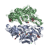
| Structure viewer | Molecule: MolmilJmol/JSmol |
|---|
- Downloads & links
-Download
| PDBx/mmCIF format | 3oe6.cif.gz | 183.5 KB | Display | PDBx/mmCIF format |
|---|---|---|---|---|
| PDB format | pdb3oe6.ent.gz | 151.8 KB | Display | PDB format |
| PDBx/mmJSON format | 3oe6.json.gz | Tree view | PDBx/mmJSON format | |
| Others |  Other downloads Other downloads |
-Validation report
| Arichive directory | https://data.pdbj.org/pub/pdb/validation_reports/oe/3oe6ftp://data.pdbj.org/pub/pdb/validation_reports/oe/3oe6 | HTTPS FTP |
|---|
-Related structure data
| Related structure data | 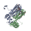 3oduC  3oe0C 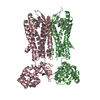 3oe8C 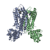 3oe9C C: citing same article ( |
|---|---|
| Similar structure data | |
| Other databases |


-Links
 PDBj
PDBj


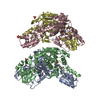
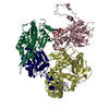
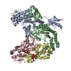
- Assembly
| Deposited unit | 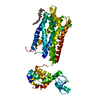
| ||||||||
|---|---|---|---|---|---|---|---|---|---|
| 1 | 
| ||||||||
| Unit cell |
|
-Components
| #1: Protein | Mass: 57537.426 Da / Num. of mol.: 1 Fragment: CXCR4 residues 2-228, LYSOZYME residues 1002-1161, CXCR4 residues 231-325 Mutation: L125W, C1054T, C1097T Source method: isolated from a genetically manipulated source Source: (gene. exp.) Homo Sapiens (human), (gene. exp.) Enterobacteria phage T4 (virus)Gene: CXCR4, CXCR4_HUMAN,E / Plasmid: pFastBac / Production host:   Spodoptera frugiperda (fall armyworm) / Strain (production host): Sf9 / References: UniProt: P61073, UniProt: P00720, lysozyme Spodoptera frugiperda (fall armyworm) / Strain (production host): Sf9 / References: UniProt: P61073, UniProt: P00720, lysozyme | ||||
|---|---|---|---|---|---|
| #2: Chemical | ChemComp-ITD / (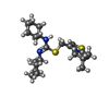  Mass: 406.651 Da / Num. of mol.: 1 / Source method: obtained synthetically / Formula: C21H34N4S2 Mass: 406.651 Da / Num. of mol.: 1 / Source method: obtained synthetically / Formula: C21H34N4S2 | ||||
| #3: Chemical |   Mass: 356.540 Da / Num. of mol.: 3 / Source method: obtained synthetically / Formula: C21H40O4 Mass: 356.540 Da / Num. of mol.: 3 / Source method: obtained synthetically / Formula: C21H40O4#4: Chemical | ChemComp-GOL / | Glycerol  Mass: 92.094 Da / Num. of mol.: 1 / Source method: obtained synthetically / Formula: C3H8O3 Mass: 92.094 Da / Num. of mol.: 1 / Source method: obtained synthetically / Formula: C3H8O3Sequence details | THE PROTEIN IS A FUSION PROTEIN WITH RESIDUES ASN1002-TYR1161 OF T4 LYSOZYME INSERTED BETWEEN ...THE PROTEIN IS A FUSION PROTEIN WITH RESIDUES ASN1002-TYR1161 OF T4 LYSOZYME INSERTED BETWEEN SER229 AND LYS230 OF CXCR4, AS INDICATED AS CXCR4-1 IN THE PUBLICATIO | |
-Experimental details
-Experiment
| Experiment | Method: X-RAY DIFFRACTION / Number of used crystals: 4 |
|---|
- Sample preparation
| Crystal | Density Matthews: 2.93 Å3/Da / Density % sol: 57.96 % |
|---|---|
| Crystal grow | Temperature: 293 K / Method: lipidic cubic phase / pH: 5.5 Details: Lipidic cubic phase made of monoolein and cholesterol, 20-26% PEG400, 0.3M Sodium malonate, 5mM Nickel chloride, 0.1M Sodium citrate pH 5.0-5.5, LIPIDIC CUBIC PHASE, temperature 293K |
-Data collection
| Diffraction | Mean temperature: 100 K |
|---|---|
| Diffraction source | Source: SYNCHROTRON / Site: APS  / Beamline: 23-ID-D / Wavelength: 1.033 Å / Beamline: 23-ID-D / Wavelength: 1.033 Å |
| Detector | Type: MARMOSAIC 300 mm CCD / Detector: CCD / Date: Feb 25, 2010 / Details: mirrors |
| Radiation | Monochromator: DOUBLE CRYSTAL / Protocol: SINGLE WAVELENGTH / Monochromatic (M) / Laue (L): M / Scattering type: x-ray |
| Radiation wavelength | Wavelength: 1.033 Å / Relative weight: 1 |
| Reflection | Resolution: 3.2→50 Å / Num. obs: 10233 / % possible obs: 94.4 % / Redundancy: 3.6 % / Biso Wilson estimate: 89.11 Å2 / Rmerge(I) obs: 0.136 / Net I/σ(I): 10.2 |
| Reflection shell | Resolution: 3.3→3.42 Å / Redundancy: 2.2 % / Rmerge(I) obs: 0.67 / Mean I/σ(I) obs: 1.5 / % possible all: 78.2 |
- Processing
| Software |
| ||||||||||||||||||||||||||||||||||||||||||||||||||||||||||||||||||||||||||||||||||||||||||||||||||||||||||||
|---|---|---|---|---|---|---|---|---|---|---|---|---|---|---|---|---|---|---|---|---|---|---|---|---|---|---|---|---|---|---|---|---|---|---|---|---|---|---|---|---|---|---|---|---|---|---|---|---|---|---|---|---|---|---|---|---|---|---|---|---|---|---|---|---|---|---|---|---|---|---|---|---|---|---|---|---|---|---|---|---|---|---|---|---|---|---|---|---|---|---|---|---|---|---|---|---|---|---|---|---|---|---|---|---|---|---|---|---|---|
| Refinement | Method to determine structure: MOLECULAR REPLACEMENT / Cor.coef. Fo:Fc: 0.8474 / Cor.coef. Fo:Fc free: 0.7467 / Highest resolution: 3.2 Å / Cross valid method: THROUGHOUT / σ(F): 0 / Stereochemistry target values: Engh & Huber
| ||||||||||||||||||||||||||||||||||||||||||||||||||||||||||||||||||||||||||||||||||||||||||||||||||||||||||||
| Displacement parameters | Biso mean: 69.12 Å2
| ||||||||||||||||||||||||||||||||||||||||||||||||||||||||||||||||||||||||||||||||||||||||||||||||||||||||||||
| Refine analyze | Luzzati coordinate error obs: 0.578 Å | ||||||||||||||||||||||||||||||||||||||||||||||||||||||||||||||||||||||||||||||||||||||||||||||||||||||||||||
| Refinement step | Cycle: LAST / Highest resolution: 3.2 Å
| ||||||||||||||||||||||||||||||||||||||||||||||||||||||||||||||||||||||||||||||||||||||||||||||||||||||||||||
| Refine LS restraints |
| ||||||||||||||||||||||||||||||||||||||||||||||||||||||||||||||||||||||||||||||||||||||||||||||||||||||||||||
| LS refinement shell | Resolution: 3.2→3.58 Å / Total num. of bins used: 5
| ||||||||||||||||||||||||||||||||||||||||||||||||||||||||||||||||||||||||||||||||||||||||||||||||||||||||||||
| Refinement TLS params. | Method: refined / Refine-ID: X-RAY DIFFRACTION
| ||||||||||||||||||||||||||||||||||||||||||||||||||||||||||||||||||||||||||||||||||||||||||||||||||||||||||||
| Refinement TLS group |
|
