 Movie
Movie Controller

[English] 日本語
 Yorodumi
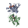
- PDB-5icw: Crystal structure of human NatF (hNaa60) homodimer bound to Coenzyme A -
+ Open data

- Basic information
| Entry | Database: PDB / ID: 5icw | |||||||||||||||||||||
|---|---|---|---|---|---|---|---|---|---|---|---|---|---|---|---|---|---|---|---|---|---|---|
| Title | Crystal structure of human NatF (hNaa60) homodimer bound to Coenzyme A | |||||||||||||||||||||
 Components Components | N-alpha-acetyltransferase 60 | |||||||||||||||||||||
 Keywords Keywords |  TRANSFERASE / Acetylation / GNAT / NAT TRANSFERASE / Acetylation / GNAT / NAT | |||||||||||||||||||||
| Function / homology |  Function and homology information Function and homology informationN-terminal methionine Nalpha-acetyltransferase NatF / N-terminal peptidyl-methionine acetylation / N-terminal protein amino acid acetylation / peptide alpha-N-acetyltransferase activity / histone H4 acetyltransferase activity / histone acetyltransferase activity / histone acetyltransferase / chromosome segregation / nucleosome assembly / cell population proliferation ...N-terminal methionine Nalpha-acetyltransferase NatF / N-terminal peptidyl-methionine acetylation / N-terminal protein amino acid acetylation / peptide alpha-N-acetyltransferase activity / histone H4 acetyltransferase activity / histone acetyltransferase activity / histone acetyltransferase / chromosome segregation / nucleosome assembly / cell population proliferation / Golgi membrane / protein homodimerization activitySimilarity search - Function | |||||||||||||||||||||
| Biological species |  Homo sapiens (human) Homo sapiens (human) | |||||||||||||||||||||
| Method | X-RAY DIFFRACTION / SYNCHROTRON / MOLECULAR REPLACEMENT / Resolution: 1.951 Å | |||||||||||||||||||||
 Authors Authors | Stove, S.I. / Magin, R.S. / Marmorstein, R. / Arnesen, T. | |||||||||||||||||||||
| Funding support |  Norway, Norway,  United States, 6items United States, 6items
| |||||||||||||||||||||
 Citation Citation | Journal: Structure / Year: 2016 Title: Crystal Structure of the Golgi-Associated Human N alpha-Acetyltransferase 60 Reveals the Molecular Determinants for Substrate-Specific Acetylation. Authors: Stve, S.I. / Magin, R.S. / Foyn, H. / Haug, B.E. / Marmorstein, R. / Arnesen, T. | |||||||||||||||||||||
| History |
|
- Structure visualization
| Structure viewer | Molecule: MolmilJmol/JSmol |
|---|
- Downloads & links
-Download
| PDBx/mmCIF format | 5icw.cif.gz | 168.3 KB | Display | PDBx/mmCIF format |
|---|---|---|---|---|
| PDB format | pdb5icw.ent.gz | 133.8 KB | Display | PDB format |
| PDBx/mmJSON format | 5icw.json.gz | Tree view | PDBx/mmJSON format | |
| Others |  Other downloads Other downloads |
-Validation report
| Arichive directory | https://data.pdbj.org/pub/pdb/validation_reports/ic/5icwftp://data.pdbj.org/pub/pdb/validation_reports/ic/5icw | HTTPS FTP |
|---|
-Related structure data
| Related structure data | 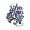 5icvC 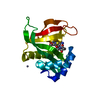 4lx9S S: Starting model for refinement C: citing same article ( |
|---|---|
| Similar structure data |


-Links
 PDBj
PDBj


- Assembly
| Deposited unit | 
| ||||||||
|---|---|---|---|---|---|---|---|---|---|
| 1 | 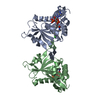
| ||||||||
| 2 | 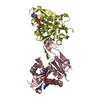
| ||||||||
| 3 | 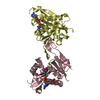
| ||||||||
| Unit cell |
|
-Components
| #1: Protein | Mass: 20767.590 Da / Num. of mol.: 4 Source method: isolated from a genetically manipulated source Source: (gene. exp.) Homo sapiens (human) / Gene: NAA60, HAT4, NAT15, UNQ2771/PRO7155 / Production host:  Escherichia coli (E. coli) Escherichia coli (E. coli)References: UniProt: Q9H7X0, histone acetyltransferase, EC: 2.3.1.88#2: Chemical | ChemComp-COA / Coenzyme A  Mass: 767.534 Da / Num. of mol.: 4 / Source method: obtained synthetically / Formula: C21H36N7O16P3S Mass: 767.534 Da / Num. of mol.: 4 / Source method: obtained synthetically / Formula: C21H36N7O16P3S#3: Chemical | ChemComp-CL / Chloride  Mass: 35.453 Da / Num. of mol.: 4 / Source method: obtained synthetically / Formula: Cl Mass: 35.453 Da / Num. of mol.: 4 / Source method: obtained synthetically / Formula: Cl#4: Water | ChemComp-HOH / | Water Mass: 18.015 Da / Num. of mol.: 464 / Source method: isolated from a natural source / Formula: H2O Mass: 18.015 Da / Num. of mol.: 464 / Source method: isolated from a natural source / Formula: H2O |
|---|
-Experimental details
-Experiment
| Experiment | Method: X-RAY DIFFRACTION / Number of used crystals: 1 |
|---|
- Sample preparation
| Crystal | Density Matthews: 2.24 Å3/Da / Density % sol: 45 % |
|---|---|
| Crystal grow | Temperature: 293.15 K / Method: vapor diffusion, hanging drop Details: 3% Tascimate, 0.1 M Bistris pH 6.5 and 16% PEG 3350 |
-Data collection
| Diffraction | Mean temperature: 100 K |
|---|---|
| Diffraction source | Source: SYNCHROTRON / Site: NSLS / Beamline: X29A / Wavelength: 1.5418 Å |
| Detector | Type: ADSC QUANTUM 315r / Detector: CCD / Date: Mar 2, 2014 |
| Radiation | Monochromator: Rigaku Osmic VariMax HF / Protocol: SINGLE WAVELENGTH / Monochromatic (M) / Laue (L): M / Scattering type: x-ray |
| Radiation wavelength | Wavelength: 1.5418 Å / Relative weight: 1 |
| Reflection | Resolution: 1.951→46.458 Å / Num. obs: 52859 / % possible obs: 94.63 % / Redundancy: 13 % / Rmerge(I) obs: 0.098 / Net I/σ(I): 12.82 |
| Reflection shell | Resolution: 1.951→1.989 Å |
- Processing
| Software |
| |||||||||||||||||||||||||||||||||||||||||||||||||||||||||||||||||||||||||||||||||||||||||||||||||||||||||||||||||||||||||||||||||||||
|---|---|---|---|---|---|---|---|---|---|---|---|---|---|---|---|---|---|---|---|---|---|---|---|---|---|---|---|---|---|---|---|---|---|---|---|---|---|---|---|---|---|---|---|---|---|---|---|---|---|---|---|---|---|---|---|---|---|---|---|---|---|---|---|---|---|---|---|---|---|---|---|---|---|---|---|---|---|---|---|---|---|---|---|---|---|---|---|---|---|---|---|---|---|---|---|---|---|---|---|---|---|---|---|---|---|---|---|---|---|---|---|---|---|---|---|---|---|---|---|---|---|---|---|---|---|---|---|---|---|---|---|---|---|---|
| Refinement | Method to determine structure: MOLECULAR REPLACEMENT Starting model: 4LX9 Resolution: 1.951→46.249 Å / SU ML: 0.25 / Cross valid method: FREE R-VALUE / σ(F): 1.37 / Phase error: 31.57 / Stereochemistry target values: ML
| |||||||||||||||||||||||||||||||||||||||||||||||||||||||||||||||||||||||||||||||||||||||||||||||||||||||||||||||||||||||||||||||||||||
| Solvent computation | Shrinkage radii: 0.9 Å / VDW probe radii: 1.11 Å / Solvent model: FLAT BULK SOLVENT MODEL | |||||||||||||||||||||||||||||||||||||||||||||||||||||||||||||||||||||||||||||||||||||||||||||||||||||||||||||||||||||||||||||||||||||
| Refinement step | Cycle: LAST / Resolution: 1.951→46.249 Å
| |||||||||||||||||||||||||||||||||||||||||||||||||||||||||||||||||||||||||||||||||||||||||||||||||||||||||||||||||||||||||||||||||||||
| Refine LS restraints |
| |||||||||||||||||||||||||||||||||||||||||||||||||||||||||||||||||||||||||||||||||||||||||||||||||||||||||||||||||||||||||||||||||||||
| LS refinement shell |
|
