 Movie
Movie Controller

[English] 日本語
 Yorodumi
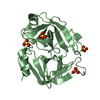
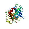
- PDB-3h7o: Crystal structure of scabies mite inactivated protease paralogue ... -
+ Open data

- Basic information
| Entry | Database: PDB / ID: 3h7o | ||||||
|---|---|---|---|---|---|---|---|
| Title | Crystal structure of scabies mite inactivated protease paralogue S-I1 (SMIPP-S-I1) | ||||||
 Components Components | Group 3 allergen SMIPP-S Yv6023A04 | ||||||
 Keywords Keywords |  HYDROLASE HYDROLASE | ||||||
| Function / homology |  Function and homology information Function and homology information | ||||||
| Biological species |  Sarcoptes scabiei type hominis (arthropod) Sarcoptes scabiei type hominis (arthropod) | ||||||
| Method | X-RAY DIFFRACTION / SYNCHROTRON / MOLECULAR REPLACEMENT / molecular replacement / Resolution: 1.85 Å | ||||||
 Authors Authors | Buckle, A.M. | ||||||
 Citation Citation | Journal: J.Mol.Biol. / Year: 2009 Title: Structural mechanisms of inactivation in scabies mite serine protease paralogues. Authors: Fischer, K. / Langendorf, C.G. / Irving, J.A. / Reynolds, S. / Willis, C. / Beckham, S. / Law, R.H. / Yang, S. / Bashtannyk-Puhalovich, T.A. / McGowan, S. / Whisstock, J.C. / Pike, R.N. / ...Authors: Fischer, K. / Langendorf, C.G. / Irving, J.A. / Reynolds, S. / Willis, C. / Beckham, S. / Law, R.H. / Yang, S. / Bashtannyk-Puhalovich, T.A. / McGowan, S. / Whisstock, J.C. / Pike, R.N. / Kemp, D.J. / Buckle, A.M. | ||||||
| History |
|
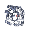
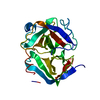
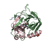
- Structure visualization
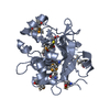
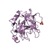
| Structure viewer | Molecule: MolmilJmol/JSmol |
|---|
- Downloads & links
-Download
| PDBx/mmCIF format | 3h7o.cif.gz | 107.9 KB | Display | PDBx/mmCIF format |
|---|---|---|---|---|
| PDB format | pdb3h7o.ent.gz | 82 KB | Display | PDB format |
| PDBx/mmJSON format | 3h7o.json.gz | Tree view | PDBx/mmJSON format | |
| Others |  Other downloads Other downloads |
-Validation report
| Arichive directory | https://data.pdbj.org/pub/pdb/validation_reports/h7/3h7oftp://data.pdbj.org/pub/pdb/validation_reports/h7/3h7o | HTTPS FTP |
|---|
-Related structure data
| Related structure data |  3h7tC  1fi8S C: citing same article ( S: Starting model for refinement |
|---|---|
| Similar structure data |


-Links
 PDBj
PDBj- Assembly
| Deposited unit | 
| ||||||||
|---|---|---|---|---|---|---|---|---|---|
| 1 | 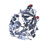
| ||||||||
| 2 | 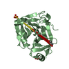
| ||||||||
| Unit cell |
|
-Components
| #1: Protein | Mass: 25263.623 Da / Num. of mol.: 2 / Fragment: UNP residues 29-256 / Mutation: N111Q, N174Q, N218Q Source method: isolated from a genetically manipulated source Details: Expression and purification as described in Sun, J. et al. (1999) Expression and purification of recombinant human granzyme B from Pichia pastoris. Biochem Biophys Res Commun 261:251-255. Source: (gene. exp.) Sarcoptes scabiei type hominis (arthropod)Plasmid: pPICZalphaA / Production host:  Pichia pastoris (fungus) / Strain (production host): KM71H / References: UniProt: Q6VPT6 Pichia pastoris (fungus) / Strain (production host): KM71H / References: UniProt: Q6VPT6#2: Chemical | ChemComp-SO4 / Sulfate  Mass: 96.063 Da / Num. of mol.: 4 / Source method: obtained synthetically / Formula: SO4 Mass: 96.063 Da / Num. of mol.: 4 / Source method: obtained synthetically / Formula: SO4#3: Chemical | ChemComp-GOL / Glycerol  Mass: 92.094 Da / Num. of mol.: 4 / Source method: obtained synthetically / Formula: C3H8O3 Mass: 92.094 Da / Num. of mol.: 4 / Source method: obtained synthetically / Formula: C3H8O3#4: Water | ChemComp-HOH / | Water Mass: 18.015 Da / Num. of mol.: 323 / Source method: isolated from a natural source / Formula: H2O Mass: 18.015 Da / Num. of mol.: 323 / Source method: isolated from a natural source / Formula: H2O |
|---|
-Experimental details
-Experiment
| Experiment | Method: X-RAY DIFFRACTION / Number of used crystals: 1 |
|---|
- Sample preparation
| Crystal | Density Matthews: 2.17 Å3/Da / Density % sol: 43.27 % |
|---|---|
| Crystal grow | Temperature: 298 K / Method: vapor diffusion, hanging drop / pH: 8.5 Details: 22% v/v PEG 8000, 10% v/v Glycerol, 0.1 M Tris-HCl pH 8.5, 0.2 M Magnesium chloride, VAPOR DIFFUSION, HANGING DROP, temperature 298K |
-Data collection
| Diffraction | Mean temperature: 100 K | ||||||||||||||||||||||||||||||||||||||||||||||||||||||||||||||||||||||||||||||||||||||||
|---|---|---|---|---|---|---|---|---|---|---|---|---|---|---|---|---|---|---|---|---|---|---|---|---|---|---|---|---|---|---|---|---|---|---|---|---|---|---|---|---|---|---|---|---|---|---|---|---|---|---|---|---|---|---|---|---|---|---|---|---|---|---|---|---|---|---|---|---|---|---|---|---|---|---|---|---|---|---|---|---|---|---|---|---|---|---|---|---|---|
| Diffraction source | Source: SYNCHROTRON / Site: APS  / Beamline: 17-ID / Wavelength: 1 Å / Beamline: 17-ID / Wavelength: 1 Å | ||||||||||||||||||||||||||||||||||||||||||||||||||||||||||||||||||||||||||||||||||||||||
| Detector | Type: ADSC QUANTUM 210 / Detector: CCD / Date: Apr 7, 2006 | ||||||||||||||||||||||||||||||||||||||||||||||||||||||||||||||||||||||||||||||||||||||||
| Radiation | Protocol: SINGLE WAVELENGTH / Monochromatic (M) / Laue (L): M / Scattering type: x-ray | ||||||||||||||||||||||||||||||||||||||||||||||||||||||||||||||||||||||||||||||||||||||||
| Radiation wavelength | Wavelength: 1 Å / Relative weight: 1 | ||||||||||||||||||||||||||||||||||||||||||||||||||||||||||||||||||||||||||||||||||||||||
| Reflection | Redundancy: 4 % / Av σ(I) over netI: 7.1 / Number: 139304 / Rmerge(I) obs: 0.067 / Rsym value: 0.067 / D res high: 1.85 Å / D res low: 65.938 Å / Num. obs: 34997 / % possible obs: 91.8 | ||||||||||||||||||||||||||||||||||||||||||||||||||||||||||||||||||||||||||||||||||||||||
| Diffraction reflection shell |
| ||||||||||||||||||||||||||||||||||||||||||||||||||||||||||||||||||||||||||||||||||||||||
| Reflection | Resolution: 1.85→57.35 Å / Num. obs: 34997 / % possible obs: 91.8 % / Redundancy: 4 % / Rmerge(I) obs: 0.067 / Rsym value: 0.067 / Net I/σ(I): 7.115 | ||||||||||||||||||||||||||||||||||||||||||||||||||||||||||||||||||||||||||||||||||||||||
| Reflection shell | Diffraction-ID: 1
|
-Phasing
| Phasing | Method: molecular replacement | |||||||||
|---|---|---|---|---|---|---|---|---|---|---|
| Phasing MR | Model details: Phaser MODE: MR_AUTO
|
- Processing
| Software |
| |||||||||||||||||||||||||||||||||||||||||||||||||||||||||||||||||||||||||||
|---|---|---|---|---|---|---|---|---|---|---|---|---|---|---|---|---|---|---|---|---|---|---|---|---|---|---|---|---|---|---|---|---|---|---|---|---|---|---|---|---|---|---|---|---|---|---|---|---|---|---|---|---|---|---|---|---|---|---|---|---|---|---|---|---|---|---|---|---|---|---|---|---|---|---|---|---|
| Refinement | Method to determine structure: MOLECULAR REPLACEMENT Starting model: PDB entry 1FI8 Resolution: 1.85→57.35 Å / Cor.coef. Fo:Fc: 0.956 / Cor.coef. Fo:Fc free: 0.941 / WRfactor Rfree: 0.222 / WRfactor Rwork: 0.182 / Occupancy max: 1 / Occupancy min: 1 / FOM work R set: 0.858 / SU B: 7.254 / SU ML: 0.097 / SU R Cruickshank DPI: 0.167 / SU Rfree: 0.146 / TLS residual ADP flag: LIKELY RESIDUAL / Cross valid method: THROUGHOUT / σ(F): 0 / ESU R: 0.168 / ESU R Free: 0.145 / Stereochemistry target values: MAXIMUM LIKELIHOOD Details: 1. HYDROGENS HAVE BEEN ADDED IN THE RIDING POSITIONS. 2. U VALUES: RESIDUAL ONLY.
| |||||||||||||||||||||||||||||||||||||||||||||||||||||||||||||||||||||||||||
| Solvent computation | Ion probe radii: 0.8 Å / Shrinkage radii: 0.8 Å / VDW probe radii: 1.4 Å / Solvent model: BABINET MODEL WITH MASK | |||||||||||||||||||||||||||||||||||||||||||||||||||||||||||||||||||||||||||
| Displacement parameters | Biso max: 74.73 Å2 / Biso mean: 31.866 Å2 / Biso min: 18.99 Å2
| |||||||||||||||||||||||||||||||||||||||||||||||||||||||||||||||||||||||||||
| Refinement step | Cycle: LAST / Resolution: 1.85→57.35 Å
| |||||||||||||||||||||||||||||||||||||||||||||||||||||||||||||||||||||||||||
| Refine LS restraints |
| |||||||||||||||||||||||||||||||||||||||||||||||||||||||||||||||||||||||||||
| LS refinement shell | Resolution: 1.85→1.898 Å / Total num. of bins used: 20
| |||||||||||||||||||||||||||||||||||||||||||||||||||||||||||||||||||||||||||
| Refinement TLS params. | Method: refined / Refine-ID: X-RAY DIFFRACTION
| |||||||||||||||||||||||||||||||||||||||||||||||||||||||||||||||||||||||||||
| Refinement TLS group |
|
