 Movie
Movie Controller

[English] 日本語
 Yorodumi
- PDB-5hyp: Structure of human C4b-binding protein alpha cain CCP domains 1 a... -
+ Open data

- Basic information
| Entry | Database: PDB / ID: 5hyp | |||||||||||||||
|---|---|---|---|---|---|---|---|---|---|---|---|---|---|---|---|---|
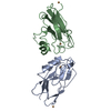
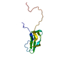
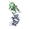
| Title | Structure of human C4b-binding protein alpha cain CCP domains 1 and 2 in complex with the hypervariable region of group A Streptococcus M28 protein | |||||||||||||||
 Components Components |
| |||||||||||||||
 Keywords Keywords |  IMMUNE SYSTEM / M protein / Complement / Streptococcus pyogenes / Hypervariable antigen IMMUNE SYSTEM / M protein / Complement / Streptococcus pyogenes / Hypervariable antigen | |||||||||||||||
| Function / homology |  Function and homology informationregulation of opsonization / response to symbiotic bacterium / negative regulation of complement activation, classical pathway / T cell mediated immunity / cell wall / complement activation, classical pathway / Regulation of Complement cascade / positive regulation of protein catabolic process / blood microparticle / innate immune response ...regulation of opsonization / response to symbiotic bacterium / negative regulation of complement activation, classical pathway / T cell mediated immunity / cell wall / complement activation, classical pathway / Regulation of Complement cascade / positive regulation of protein catabolic process / blood microparticle / innate immune response / extracellular space / RNA binding / extracellular region / plasma membrane Function and homology informationregulation of opsonization / response to symbiotic bacterium / negative regulation of complement activation, classical pathway / T cell mediated immunity / cell wall / complement activation, classical pathway / Regulation of Complement cascade / positive regulation of protein catabolic process / blood microparticle / innate immune response ...regulation of opsonization / response to symbiotic bacterium / negative regulation of complement activation, classical pathway / T cell mediated immunity / cell wall / complement activation, classical pathway / Regulation of Complement cascade / positive regulation of protein catabolic process / blood microparticle / innate immune response / extracellular space / RNA binding / extracellular region / plasma membraneSimilarity search - Function | |||||||||||||||
| Biological species |  Homo sapiens (human) Homo sapiens (human) Streptococcus pyogenes (bacteria) Streptococcus pyogenes (bacteria) | |||||||||||||||
| Method | X-RAY DIFFRACTION / SYNCHROTRON / MOLECULAR REPLACEMENT / Resolution: 3.024 Å | |||||||||||||||
 Authors Authors | Buffalo, C.Z. / Bahn-Suh, A.J. / Ghosh, P. | |||||||||||||||
| Funding support |  United States, 4items United States, 4items
| |||||||||||||||
 Citation Citation | Journal: Nat Microbiol / Year: 2016 Title: Conserved patterns hidden within group A Streptococcus M protein hypervariability recognize human C4b-binding protein. Authors: Buffalo, C.Z. / Bahn-Suh, A.J. / Hirakis, S.P. / Biswas, T. / Amaro, R.E. / Nizet, V. / Ghosh, P. | |||||||||||||||
| History |
|
- Structure visualization
| Structure viewer | Molecule: MolmilJmol/JSmol |
|---|
- Downloads & links
-Download
| PDBx/mmCIF format | 5hyp.cif.gz | 79.5 KB | Display | PDBx/mmCIF format |
|---|---|---|---|---|
| PDB format | pdb5hyp.ent.gz | 59.2 KB | Display | PDB format |
| PDBx/mmJSON format | 5hyp.json.gz | Tree view | PDBx/mmJSON format | |
| Others |  Other downloads Other downloads |
-Validation report
| Arichive directory | https://data.pdbj.org/pub/pdb/validation_reports/hy/5hypftp://data.pdbj.org/pub/pdb/validation_reports/hy/5hyp | HTTPS FTP |
|---|
-Related structure data
| Related structure data |  5hytC 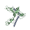 5hyuSC 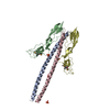 5hzpC 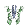 5i0qC S: Starting model for refinement C: citing same article ( |
|---|---|
| Similar structure data |
-Links
 PDBj
PDBj

- Assembly
| Deposited unit | 
| ||||||||
|---|---|---|---|---|---|---|---|---|---|
| 1 |
| ||||||||
| Unit cell |
|
-Components
| #1: Protein | Mass: 14163.894 Da / Num. of mol.: 1 / Fragment: UNP residues 49-172 Source method: isolated from a genetically manipulated source Source: (gene. exp.) Homo sapiens (human) / Gene: C4BPA, C4BP / Production host: Escherichia coli (E. coli) / References: UniProt: P04003 |
|---|---|
| #2: Protein | Mass: 12193.425 Da / Num. of mol.: 1 / Fragment: UNP residues 42-141 Source method: isolated from a genetically manipulated source Source: (gene. exp.) Streptococcus pyogenes (bacteria) / Gene: emm28 / Production host: Escherichia coli (E. coli) / References: UniProt: W0T1Y4 |
-Experimental details
-Experiment
| Experiment | Method: X-RAY DIFFRACTION / Number of used crystals: 1 |
|---|
- Sample preparation
| Crystal | Density Matthews: 3.74 Å3/Da / Density % sol: 67.07 % |
|---|---|
| Crystal grow | Temperature: 293 K / Method: vapor diffusion / pH: 7 / Details: 1.5 M Ammonium Sulfate, 0.1 M Bis-Tris Propane |
-Data collection
| Diffraction | Mean temperature: 100 K |
|---|---|
| Diffraction source | Source: SYNCHROTRON / Site: ALS / Beamline: 8.2.1 / Wavelength: 0.979 Å |
| Detector | Type: ADSC QUANTUM 315r / Detector: CCD / Date: Apr 14, 2014 |
| Radiation | Protocol: SINGLE WAVELENGTH / Monochromatic (M) / Laue (L): M / Scattering type: x-ray |
| Radiation wavelength | Wavelength: 0.979 Å / Relative weight: 1 |
| Reflection | Resolution: 3.02→44.397 Å / Num. obs: 8361 / % possible obs: 99.8 % / Redundancy: 9.5 % / CC1/2: 0.48 / Rmerge(I) obs: 0.14 / Net I/σ(I): 13.2 |
| Reflection shell | Resolution: 3.02→3.07 Å / Redundancy: 9.7 % / Rmerge(I) obs: 1 / Mean I/σ(I) obs: 0.81 / % possible all: 100 |
- Processing
| Software |
| |||||||||||||||||||||||||||||||||||||||||||||||||||||||||||||||||||||||||||||||||||||||||||||||||||||||||||||||||||||||||||||
|---|---|---|---|---|---|---|---|---|---|---|---|---|---|---|---|---|---|---|---|---|---|---|---|---|---|---|---|---|---|---|---|---|---|---|---|---|---|---|---|---|---|---|---|---|---|---|---|---|---|---|---|---|---|---|---|---|---|---|---|---|---|---|---|---|---|---|---|---|---|---|---|---|---|---|---|---|---|---|---|---|---|---|---|---|---|---|---|---|---|---|---|---|---|---|---|---|---|---|---|---|---|---|---|---|---|---|---|---|---|---|---|---|---|---|---|---|---|---|---|---|---|---|---|---|---|---|
| Refinement | Method to determine structure: MOLECULAR REPLACEMENT Starting model: 5HYU Resolution: 3.024→44.397 Å / SU ML: 0.42 / Cross valid method: FREE R-VALUE / σ(F): 0 / Phase error: 31.02 / Stereochemistry target values: ML
| |||||||||||||||||||||||||||||||||||||||||||||||||||||||||||||||||||||||||||||||||||||||||||||||||||||||||||||||||||||||||||||
| Solvent computation | Shrinkage radii: 0.9 Å / VDW probe radii: 1.11 Å / Solvent model: FLAT BULK SOLVENT MODEL | |||||||||||||||||||||||||||||||||||||||||||||||||||||||||||||||||||||||||||||||||||||||||||||||||||||||||||||||||||||||||||||
| Refinement step | Cycle: LAST / Resolution: 3.024→44.397 Å
| |||||||||||||||||||||||||||||||||||||||||||||||||||||||||||||||||||||||||||||||||||||||||||||||||||||||||||||||||||||||||||||
| Refine LS restraints |
| |||||||||||||||||||||||||||||||||||||||||||||||||||||||||||||||||||||||||||||||||||||||||||||||||||||||||||||||||||||||||||||
| LS refinement shell |
| |||||||||||||||||||||||||||||||||||||||||||||||||||||||||||||||||||||||||||||||||||||||||||||||||||||||||||||||||||||||||||||
| Refinement TLS params. | Method: refined / Refine-ID: X-RAY DIFFRACTION
| |||||||||||||||||||||||||||||||||||||||||||||||||||||||||||||||||||||||||||||||||||||||||||||||||||||||||||||||||||||||||||||
| Refinement TLS group |
|
