 Movie
Movie Controller

[English] 日本語
 Yorodumi
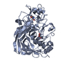
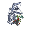
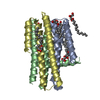
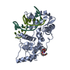
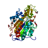
- PDB-5hnf: Crystal structure of pyrene- and phenanthrene-modified DNA in com... -
+ Open data

- Basic information
| Entry | Database: PDB / ID: 5hnf | ||||||
|---|---|---|---|---|---|---|---|
| Title | Crystal structure of pyrene- and phenanthrene-modified DNA in complex with the BpuJ1 endonuclease binding domain | ||||||
 Components Components |
| ||||||
 Keywords Keywords |  HYDROLASE / Phenanthrene / Pyrene / DNA / Endonuclease HYDROLASE / Phenanthrene / Pyrene / DNA / Endonuclease | ||||||
| Function / homology |  Function and homology information Function and homology information | ||||||
| Biological species |  Bacillus pumilus (bacteria) Bacillus pumilus (bacteria)synthetic construct (others) | ||||||
| Method | X-RAY DIFFRACTION / SYNCHROTRON / MOLECULAR REPLACEMENT / Resolution: 1.546 Å | ||||||
 Authors Authors | Probst, M. / Aeschimann, W. / Chau, T.-T.-H. / Langenegger, S.M. / Stocker, A. / Haener, R. | ||||||
 Citation Citation | Journal: Nucleic Acids Res. / Year: 2016 Title: Structural insight into DNA-assembled oligochromophores: crystallographic analysis of pyrene- and phenanthrene-modified DNA in complex with BpuJI endonuclease. Authors: Probst, M. / Aeschimann, W. / Chau, T.T. / Langenegger, S.M. / Stocker, A. / Haner, R. | ||||||
| History |
|
- Structure visualization
| Structure viewer | Molecule: MolmilJmol/JSmol |
|---|
- Downloads & links
-Download
| PDBx/mmCIF format | 5hnf.cif.gz | 167.4 KB | Display | PDBx/mmCIF format |
|---|---|---|---|---|
| PDB format | pdb5hnf.ent.gz | 128.1 KB | Display | PDB format |
| PDBx/mmJSON format | 5hnf.json.gz | Tree view | PDBx/mmJSON format | |
| Others |  Other downloads Other downloads |
-Validation report
| Arichive directory | https://data.pdbj.org/pub/pdb/validation_reports/hn/5hnfftp://data.pdbj.org/pub/pdb/validation_reports/hn/5hnf | HTTPS FTP |
|---|
-Related structure data
| Related structure data |  5hltC  5hnhC  2vlaS S: Starting model for refinement C: citing same article ( |
|---|---|
| Similar structure data |


-Links
 PDBj
PDBj


- Assembly
| Deposited unit | 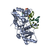
| ||||||||
|---|---|---|---|---|---|---|---|---|---|
| 1 |
| ||||||||
| Unit cell |
| ||||||||
| Components on special symmetry positions |
|
-Components
| #1: Protein | Mass: 33766.219 Da / Num. of mol.: 1 Source method: isolated from a genetically manipulated source Source: (gene. exp.) Bacillus pumilus (bacteria) / Gene: bpuJIR / Production host: Escherichia coli (E. coli) / References: UniProt: A3FMN7 |
|---|---|
| #2: DNA chain | Mass: 3470.380 Da / Num. of mol.: 1 / Source method: obtained synthetically / Source: (synth.) synthetic construct (others) |
| #3: DNA chain | Mass: 3397.321 Da / Num. of mol.: 1 / Source method: obtained synthetically / Source: (synth.) synthetic construct (others) |
| #4: Water | ChemComp-HOH / Water Mass: 18.015 Da / Num. of mol.: 479 / Source method: isolated from a natural source / Formula: H2O Mass: 18.015 Da / Num. of mol.: 479 / Source method: isolated from a natural source / Formula: H2O |
-Experimental details
-Experiment
| Experiment | Method: X-RAY DIFFRACTION / Number of used crystals: 1 |
|---|
- Sample preparation
| Crystal | Density Matthews: 2.69 Å3/Da / Density % sol: 54.22 % |
|---|---|
| Crystal grow | Temperature: 291 K / Method: vapor diffusion, sitting drop / pH: 7 / Details: Ammonium citrate, PEG 3350 |
-Data collection
| Diffraction | Mean temperature: 100 K |
|---|---|
| Diffraction source | Source: SYNCHROTRON / Site: SLS  / Beamline: X06DA / Wavelength: 1.00004 Å / Beamline: X06DA / Wavelength: 1.00004 Å |
| Detector | Type: DECTRIS PILATUS 2M / Detector: PIXEL / Date: Jun 10, 2015 |
| Radiation | Protocol: SINGLE WAVELENGTH / Monochromatic (M) / Laue (L): M / Scattering type: x-ray |
| Radiation wavelength | Wavelength: 1.00004 Å / Relative weight: 1 |
| Reflection | Resolution: 1.546→43.93 Å / Num. obs: 61076 / % possible obs: 98 % / Redundancy: 3.17 % / CC1/2: 0.999 / Rmerge(I) obs: 0.046 / Net I/σ(I): 16.7 |
| Reflection shell | Resolution: 1.55→1.64 Å / Redundancy: 3.12 % / Rmerge(I) obs: 0.375 / Mean I/σ(I) obs: 3.03 / % possible all: 95.1 |
- Processing
| Software |
| |||||||||||||||||||||||||||||||||||||||||||||||||||||||||||||||||||||||||||||||||||||||||||||||||||||||||||||||||||||||||||||||||||||||||||||||||||||||||||||||||||||||||||||||||||||||||||||||||||||||||||||||||||||||||
|---|---|---|---|---|---|---|---|---|---|---|---|---|---|---|---|---|---|---|---|---|---|---|---|---|---|---|---|---|---|---|---|---|---|---|---|---|---|---|---|---|---|---|---|---|---|---|---|---|---|---|---|---|---|---|---|---|---|---|---|---|---|---|---|---|---|---|---|---|---|---|---|---|---|---|---|---|---|---|---|---|---|---|---|---|---|---|---|---|---|---|---|---|---|---|---|---|---|---|---|---|---|---|---|---|---|---|---|---|---|---|---|---|---|---|---|---|---|---|---|---|---|---|---|---|---|---|---|---|---|---|---|---|---|---|---|---|---|---|---|---|---|---|---|---|---|---|---|---|---|---|---|---|---|---|---|---|---|---|---|---|---|---|---|---|---|---|---|---|---|---|---|---|---|---|---|---|---|---|---|---|---|---|---|---|---|---|---|---|---|---|---|---|---|---|---|---|---|---|---|---|---|---|---|---|---|---|---|---|---|---|---|---|---|---|---|---|---|---|
| Refinement | Method to determine structure: MOLECULAR REPLACEMENT Starting model: 2VLA Resolution: 1.546→43.93 Å / SU ML: 0.16 / Cross valid method: FREE R-VALUE / σ(F): 0.1 / Phase error: 19.44 / Stereochemistry target values: ML
| |||||||||||||||||||||||||||||||||||||||||||||||||||||||||||||||||||||||||||||||||||||||||||||||||||||||||||||||||||||||||||||||||||||||||||||||||||||||||||||||||||||||||||||||||||||||||||||||||||||||||||||||||||||||||
| Solvent computation | Shrinkage radii: 0.9 Å / VDW probe radii: 1.11 Å / Solvent model: FLAT BULK SOLVENT MODEL | |||||||||||||||||||||||||||||||||||||||||||||||||||||||||||||||||||||||||||||||||||||||||||||||||||||||||||||||||||||||||||||||||||||||||||||||||||||||||||||||||||||||||||||||||||||||||||||||||||||||||||||||||||||||||
| Refinement step | Cycle: LAST / Resolution: 1.546→43.93 Å
| |||||||||||||||||||||||||||||||||||||||||||||||||||||||||||||||||||||||||||||||||||||||||||||||||||||||||||||||||||||||||||||||||||||||||||||||||||||||||||||||||||||||||||||||||||||||||||||||||||||||||||||||||||||||||
| Refine LS restraints |
| |||||||||||||||||||||||||||||||||||||||||||||||||||||||||||||||||||||||||||||||||||||||||||||||||||||||||||||||||||||||||||||||||||||||||||||||||||||||||||||||||||||||||||||||||||||||||||||||||||||||||||||||||||||||||
| LS refinement shell |
| |||||||||||||||||||||||||||||||||||||||||||||||||||||||||||||||||||||||||||||||||||||||||||||||||||||||||||||||||||||||||||||||||||||||||||||||||||||||||||||||||||||||||||||||||||||||||||||||||||||||||||||||||||||||||
| Refinement TLS params. | Method: refined / Refine-ID: X-RAY DIFFRACTION
| |||||||||||||||||||||||||||||||||||||||||||||||||||||||||||||||||||||||||||||||||||||||||||||||||||||||||||||||||||||||||||||||||||||||||||||||||||||||||||||||||||||||||||||||||||||||||||||||||||||||||||||||||||||||||
| Refinement TLS group |
|
