 Movie
Movie Controller


+ Open data

- Basic information
| Entry | Database: PDB / ID: 5hkh | ||||||
|---|---|---|---|---|---|---|---|
| Title | Crystal structure of Ufm1 in complex with UBA5 | ||||||
 Components Components |
| ||||||
 Keywords Keywords |  SIGNALING PROTEIN SIGNALING PROTEIN | ||||||
| Function / homology |  Function and homology information Function and homology informationUFM1 activating enzyme activity / protein ufmylation / protein K69-linked ufmylation / megakaryocyte differentiation / regulation of intracellular estrogen receptor signaling pathway / reticulophagy / neuromuscular process / negative regulation of protein import into nucleus / localization / response to endoplasmic reticulum stress ...UFM1 activating enzyme activity / protein ufmylation / protein K69-linked ufmylation / megakaryocyte differentiation / regulation of intracellular estrogen receptor signaling pathway / reticulophagy / neuromuscular process / negative regulation of protein import into nucleus / localization / response to endoplasmic reticulum stress / erythrocyte differentiation / brain development / Antigen processing: Ubiquitination & Proteasome degradation / intracellular membrane-bounded organelle / endoplasmic reticulum membrane / negative regulation of apoptotic process / Golgi apparatus / endoplasmic reticulum / protein homodimerization activity / zinc ion binding / ATP binding / nucleus / cytosol / cytoplasmSimilarity search - Function | ||||||
| Biological species |  Homo sapiens (human) Homo sapiens (human) | ||||||
| Method | X-RAY DIFFRACTION / SYNCHROTRON / MOLECULAR REPLACEMENT / Resolution: 2.55 Å | ||||||
 Authors Authors | Huber, J. / Doetsch, V. / Rogov, V.V. / Akutsu, M. | ||||||
 Citation Citation | Journal: J.Biol.Chem. / Year: 2016 Title: Structural and Functional Analysis of a Novel Interaction Motif within UFM1-activating Enzyme 5 (UBA5) Required for Binding to Ubiquitin-like Proteins and Ufmylation. Authors: Habisov, S. / Huber, J. / Ichimura, Y. / Akutsu, M. / Rogova, N. / Loehr, F. / McEwan, D.G. / Johansen, T. / Dikic, I. / Doetsch, V. / Komatsu, M. / Rogov, V.V. / Kirkin, V. | ||||||
| History |
|
- Structure visualization
| Structure viewer | Molecule: MolmilJmol/JSmol |
|---|
- Downloads & links
-Download
| PDBx/mmCIF format | 5hkh.cif.gz | 45.8 KB | Display | PDBx/mmCIF format |
|---|---|---|---|---|
| PDB format | pdb5hkh.ent.gz | 31.8 KB | Display | PDB format |
| PDBx/mmJSON format | 5hkh.json.gz | Tree view | PDBx/mmJSON format | |
| Others |  Other downloads Other downloads |
-Validation report
| Arichive directory | https://data.pdbj.org/pub/pdb/validation_reports/hk/5hkhftp://data.pdbj.org/pub/pdb/validation_reports/hk/5hkh | HTTPS FTP |
|---|
-Related structure data
| Related structure data | 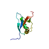 1wxsS S: Starting model for refinement |
|---|---|
| Similar structure data |


-Links
 PDBj
PDBj

- Assembly
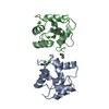
| Deposited unit | 
| ||||||||
|---|---|---|---|---|---|---|---|---|---|
| 1 |
| ||||||||
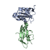
| Unit cell |
| ||||||||
| Components on special symmetry positions |
|
-Components
| #1: Protein | Mass: 9195.574 Da / Num. of mol.: 2 Source method: isolated from a genetically manipulated source Source: (gene. exp.) Homo sapiens (human) / Gene: UFM1, C13orf20, BM-002 / Production host:  Escherichia coli (E. coli) / References: UniProt: P61960 Escherichia coli (E. coli) / References: UniProt: P61960#2: Protein/peptide | | Mass: 1074.141 Da / Num. of mol.: 1 / Source method: obtained synthetically / Source: (synth.) Homo sapiens (human) / References: UniProt: Q9GZZ9*PLUS#3: Water | ChemComp-HOH / | Water Mass: 18.015 Da / Num. of mol.: 25 / Source method: isolated from a natural source / Formula: H2O Mass: 18.015 Da / Num. of mol.: 25 / Source method: isolated from a natural source / Formula: H2O |
|---|
-Experimental details
-Experiment
| Experiment | Method: X-RAY DIFFRACTION / Number of used crystals: 1 |
|---|
- Sample preparation
| Crystal | Density Matthews: 3.14 Å3/Da / Density % sol: 60.86 % |
|---|---|
| Crystal grow | Temperature: 293 K / Method: vapor diffusion, sitting drop Details: 0.2 M ammonium acetate, 0.01 M magnesium acetate, 30% polyethylene glycol 8000, 0.05 M sodium cacodylate, pH 8.0 |
-Data collection
| Diffraction | Mean temperature: 77 K |
|---|---|
| Diffraction source | Source: SYNCHROTRON / Site: SLS  / Beamline: X06DA / Wavelength: 1.072 Å / Beamline: X06DA / Wavelength: 1.072 Å |
| Detector | Type: DECTRIS PILATUS 2M / Detector: PIXEL / Date: Dec 12, 2014 |
| Radiation | Protocol: SINGLE WAVELENGTH / Monochromatic (M) / Laue (L): M / Scattering type: x-ray |
| Radiation wavelength | Wavelength: 1.072 Å / Relative weight: 1 |
| Reflection | Resolution: 2.55→46.88 Å / Num. obs: 8280 / % possible obs: 100 % / Redundancy: 19.4 % / Net I/σ(I): 41.5 |
- Processing
| Software |
| ||||||||||||||||||||||||||||||||||||||||||
|---|---|---|---|---|---|---|---|---|---|---|---|---|---|---|---|---|---|---|---|---|---|---|---|---|---|---|---|---|---|---|---|---|---|---|---|---|---|---|---|---|---|---|---|
| Refinement | Method to determine structure: MOLECULAR REPLACEMENT Starting model: 1WXS Resolution: 2.55→46.877 Å / SU ML: 0.25 / Cross valid method: FREE R-VALUE / σ(F): 1.34 / Phase error: 25.08
| ||||||||||||||||||||||||||||||||||||||||||
| Solvent computation | Shrinkage radii: 0.9 Å / VDW probe radii: 1.11 Å | ||||||||||||||||||||||||||||||||||||||||||
| Displacement parameters | Biso max: 85.39 Å2 / Biso mean: 47.31 Å2 / Biso min: 28.33 Å2 | ||||||||||||||||||||||||||||||||||||||||||
| Refinement step | Cycle: final / Resolution: 2.55→46.877 Å
| ||||||||||||||||||||||||||||||||||||||||||
| Refine LS restraints |
| ||||||||||||||||||||||||||||||||||||||||||
| LS refinement shell | Refine-ID: X-RAY DIFFRACTION / Total num. of bins used: 6 / % reflection obs: 100 %
|
