 Movie
Movie Controller

[English] 日本語
 Yorodumi
- PDB-3g1j: Structure from the mobile metagenome of Vibrio cholerae. Integron... -
+ Open data

- Basic information
| Entry | Database: PDB / ID: 3g1j | ||||||
|---|---|---|---|---|---|---|---|
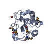
| Title | Structure from the mobile metagenome of Vibrio cholerae. Integron cassette protein VCH_CASS4. | ||||||
 Components Components | Integron cassette protein | ||||||
 Keywords Keywords |  STRUCTURAL GENOMICS / UNKNOWN FUNCTION / Novel / Integron cassette protein / Vibrio cholerae / Oyster pond / Woodshole / USA / PSI-2 / Protein Structure Initiative / Midwest Center for Structural Genomics / MCSG STRUCTURAL GENOMICS / UNKNOWN FUNCTION / Novel / Integron cassette protein / Vibrio cholerae / Oyster pond / Woodshole / USA / PSI-2 / Protein Structure Initiative / Midwest Center for Structural Genomics / MCSG | ||||||
| Function / homology | mobile metagenome of vibrio cholerae. Integron cassette protein vch_cass4. / Domain of unknown function DUF3601 / Domain of unknown function (DUF3601) / SH3 type barrels. / Roll / Mainly Beta / Integron cassette protein Function and homology information Function and homology information | ||||||
| Biological species |   Vibrio cholerae (bacteria) Vibrio cholerae (bacteria) | ||||||
| Method | X-RAY DIFFRACTION / SYNCHROTRON / SAD / Resolution: 1.7 Å | ||||||
 Authors Authors | Deshpande, C.N. / Sureshan, V. / Harrop, S.J. / Boucher, Y. / Xu, X. / Cui, H. / Edwards, A. / Savchenko, A. / Joachimiak, A. / Tan, K. ...Deshpande, C.N. / Sureshan, V. / Harrop, S.J. / Boucher, Y. / Xu, X. / Cui, H. / Edwards, A. / Savchenko, A. / Joachimiak, A. / Tan, K. / Stokes, H.W. / Curmi, P.M.G. / Mabbutt, B.C. / Midwest Center for Structural Genomics (MCSG) | ||||||
 Citation Citation | Journal: To be published Title: Structure from the mobile metagenome of Vibrio cholerae. Integron cassette protein VCH_CASS4. Authors: Deshpande, C.N. / Sureshan, V. / Harrop, S.J. / Boucher, Y. / Xu, X. / Cui, H. / Edwards, A. / Savchenko, A. / Joachimiak, A. / Tan, K. / Stokes, H.W. / Curmi, P.M.G. / Mabbutt, B.C. | ||||||
| History |
|
- Structure visualization
| Structure viewer | Molecule: MolmilJmol/JSmol |
|---|
- Downloads & links
-Download
| PDBx/mmCIF format | 3g1j.cif.gz | 86 KB | Display | PDBx/mmCIF format |
|---|---|---|---|---|
| PDB format | pdb3g1j.ent.gz | 71 KB | Display | PDB format |
| PDBx/mmJSON format | 3g1j.json.gz | Tree view | PDBx/mmJSON format | |
| Others |  Other downloads Other downloads |
-Validation report
| Arichive directory | https://data.pdbj.org/pub/pdb/validation_reports/g1/3g1jftp://data.pdbj.org/pub/pdb/validation_reports/g1/3g1j | HTTPS FTP |
|---|
-Related structure data
| Similar structure data | |
|---|---|
| Other databases |


-Links
 PDBj
PDBj- Assembly
| Deposited unit | 
| |||||||||
|---|---|---|---|---|---|---|---|---|---|---|
| 1 | 
| |||||||||
| 2 | 
| |||||||||
| Unit cell |
| |||||||||
| Components on special symmetry positions |
|
-Components
| #1: Protein | Mass: 11147.068 Da / Num. of mol.: 2 Source method: isolated from a genetically manipulated source Source: (gene. exp.) Vibrio cholerae (bacteria) / Plasmid: p15TV-L / Production host: Escherichia coli (E. coli) / References: UniProt: D0VX16*PLUS#2: Water | ChemComp-HOH / | Water Mass: 18.015 Da / Num. of mol.: 219 / Source method: isolated from a natural source / Formula: H2O Mass: 18.015 Da / Num. of mol.: 219 / Source method: isolated from a natural source / Formula: H2OSequence details | A SEQUENCE DATABASE REFERENCE FOR THIS PROTEIN DOES NOT CURRENTLY EXIST IN UNIPROT | |
|---|
-Experimental details
-Experiment
| Experiment | Method: X-RAY DIFFRACTION / Number of used crystals: 1 |
|---|
- Sample preparation
| Crystal | Density Matthews: 2.56 Å3/Da / Density % sol: 52 % |
|---|---|
| Crystal grow | Temperature: 296 K / Method: vapor diffusion, sitting drop / pH: 4.6 Details: 0.1M Na(OAC), 0.2M NaFormate, pH 4.6, VAPOR DIFFUSION, SITTING DROP, temperature 296K |
-Data collection
| Diffraction | Mean temperature: 100 K |
|---|---|
| Diffraction source | Source: SYNCHROTRON / Site: APS  / Beamline: 19-BM / Wavelength: 0.97942 Å / Beamline: 19-BM / Wavelength: 0.97942 Å |
| Detector | Type: SBC-3 / Detector: CCD / Date: Oct 24, 2008 |
| Radiation | Protocol: SINGLE WAVELENGTH / Monochromatic (M) / Laue (L): M / Scattering type: x-ray |
| Radiation wavelength | Wavelength: 0.97942 Å / Relative weight: 1 |
| Reflection | Resolution: 1.7→24.668 Å / Num. all: 25217 / Num. obs: 25217 / % possible obs: 91.7 % / Observed criterion σ(F): 0 / Observed criterion σ(I): 0 / Redundancy: 6.4 % / Biso Wilson estimate: 28.65 Å2 / Rmerge(I) obs: 0.041 / Rsym value: 0.041 / Net I/σ(I): 37.14 |
| Reflection shell | Resolution: 1.7→1.73 Å / Redundancy: 5.1 % / Rmerge(I) obs: 0.615 / Mean I/σ(I) obs: 2.2 / Num. unique all: 1234 / Rsym value: 0.615 / % possible all: 100 |
- Processing
| Software |
| ||||||||||||||||||||||||||||||||||||||||||||||||||||||||||||||||||||||||||||||||||||
|---|---|---|---|---|---|---|---|---|---|---|---|---|---|---|---|---|---|---|---|---|---|---|---|---|---|---|---|---|---|---|---|---|---|---|---|---|---|---|---|---|---|---|---|---|---|---|---|---|---|---|---|---|---|---|---|---|---|---|---|---|---|---|---|---|---|---|---|---|---|---|---|---|---|---|---|---|---|---|---|---|---|---|---|---|---|
| Refinement | Method to determine structure: SAD / Resolution: 1.7→24.66 Å / Occupancy max: 1 / Occupancy min: 0.33 / SU ML: 0.24 / Isotropic thermal model: Isotropic / Cross valid method: THROUGHOUT / σ(F): 0.05 / σ(I): 0 / Phase error: 21.65 / Stereochemistry target values: MLHL
| ||||||||||||||||||||||||||||||||||||||||||||||||||||||||||||||||||||||||||||||||||||
| Solvent computation | Shrinkage radii: 0.9 Å / VDW probe radii: 1.11 Å / Solvent model: FLAT BULK SOLVENT MODEL / Bsol: 68.391 Å2 / ksol: 0.382 e/Å3 | ||||||||||||||||||||||||||||||||||||||||||||||||||||||||||||||||||||||||||||||||||||
| Displacement parameters | Biso max: 133.6 Å2 / Biso min: 7.86 Å2
| ||||||||||||||||||||||||||||||||||||||||||||||||||||||||||||||||||||||||||||||||||||
| Refinement step | Cycle: LAST / Resolution: 1.7→24.66 Å
| ||||||||||||||||||||||||||||||||||||||||||||||||||||||||||||||||||||||||||||||||||||
| Refine LS restraints |
| ||||||||||||||||||||||||||||||||||||||||||||||||||||||||||||||||||||||||||||||||||||
| LS refinement shell | Refine-ID: X-RAY DIFFRACTION / Total num. of bins used: 13
| ||||||||||||||||||||||||||||||||||||||||||||||||||||||||||||||||||||||||||||||||||||
| Refinement TLS params. | Method: refined / Refine-ID: X-RAY DIFFRACTION
| ||||||||||||||||||||||||||||||||||||||||||||||||||||||||||||||||||||||||||||||||||||
| Refinement TLS group |
|
