 Movie
Movie Controller


+ Open data

- Basic information
| Entry | Database: PDB / ID: 5hew | ||||||
|---|---|---|---|---|---|---|---|
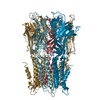
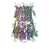
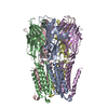
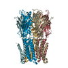
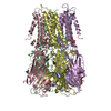
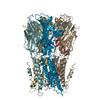
| Title | Pentameric ligand-gated ion channel ELIC mutant T28D | ||||||
 Components Components | Gamma-aminobutyric-acid receptor subunit beta-1 | ||||||
 Keywords Keywords |  TRANSPORT PROTEIN / Pentameric ligand-gated ion channels / Membrane protein / Ligand-gated ion channel TRANSPORT PROTEIN / Pentameric ligand-gated ion channels / Membrane protein / Ligand-gated ion channel | ||||||
| Function / homology |  Function and homology information Function and homology informationextracellular ligand-gated monoatomic ion channel activity / transmembrane signaling receptor activity / membrane / identical protein bindingSimilarity search - Function | ||||||
| Biological species |  Dickeya dadantii (bacteria) Dickeya dadantii (bacteria) | ||||||
| Method | X-RAY DIFFRACTION / SYNCHROTRON / MOLECULAR REPLACEMENT / Resolution: 4.5 Å | ||||||
 Authors Authors | Engeler, S. / Dutzler, R. | ||||||
| Funding support |  Switzerland, 1items Switzerland, 1items
| ||||||
 Citation Citation | Journal: Plos Biol. / Year: 2016 Title: Signal Transduction at the Domain Interface of Prokaryotic Pentameric Ligand-Gated Ion Channels. Authors: Bertozzi, C. / Zimmermann, I. / Engeler, S. / Hilf, R.J. / Dutzler, R. | ||||||
| History |
|
- Structure visualization
| Structure viewer | Molecule: MolmilJmol/JSmol |
|---|
- Downloads & links
-Download
| PDBx/mmCIF format | 5hew.cif.gz | 607.1 KB | Display | PDBx/mmCIF format |
|---|---|---|---|---|
| PDB format | pdb5hew.ent.gz | 507.3 KB | Display | PDB format |
| PDBx/mmJSON format | 5hew.json.gz | Tree view | PDBx/mmJSON format | |
| Others |  Other downloads Other downloads |
-Validation report
| Arichive directory | https://data.pdbj.org/pub/pdb/validation_reports/he/5hewftp://data.pdbj.org/pub/pdb/validation_reports/he/5hew | HTTPS FTP |
|---|
-Related structure data
| Related structure data | 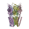 5hegC  5hehC  5hejC  5heoC  5heuC  2yn6S S: Starting model for refinement C: citing same article ( |
|---|---|
| Similar structure data |

-Links
 PDBj
PDBj

- Assembly
| Deposited unit | 
| ||||||||
|---|---|---|---|---|---|---|---|---|---|
| 1 | 
| ||||||||
| 2 | 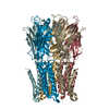
| ||||||||
| Unit cell |
|
-Components
| #1: Protein | Mass: 36892.984 Da / Num. of mol.: 10 Source method: isolated from a genetically manipulated source Source: (gene. exp.) Dickeya dadantii (strain 3937) (bacteria)Strain: 3937 / Gene: Dda3937_00520 / Production host: Escherichia coli (E. coli) / References: UniProt: E0SJQ4 |
|---|
-Experimental details
-Experiment
| Experiment | Method: X-RAY DIFFRACTION |
|---|
- Sample preparation
| Crystal | Density Matthews: 3.94 Å3/Da / Density % sol: 68.77 % |
|---|---|
| Crystal grow | Temperature: 277 K / Method: vapor diffusion, sitting drop / pH: 6.5 / Details: PEG 4000 ammonium sulfate |
-Data collection
| Diffraction | Mean temperature: 100 K |
|---|---|
| Diffraction source | Source: SYNCHROTRON / Site: SLS / Beamline: X06SA / Wavelength: 1.603 Å |
| Detector | Type: DECTRIS PILATUS 6M / Detector: PIXEL / Date: Apr 16, 2015 |
| Radiation | Protocol: SINGLE WAVELENGTH / Monochromatic (M) / Laue (L): M / Scattering type: x-ray |
| Radiation wavelength | Wavelength: 1.603 Å / Relative weight: 1 |
| Reflection | Resolution: 4.5→50 Å / Num. obs: 33795 / % possible obs: 99.7 % / Redundancy: 11.9 % / Rmerge(I) obs: 0.093 / Net I/σ(I): 13.1 |
| Reflection shell | Resolution: 4.5→4.6 Å / Redundancy: 1.6 % / Rmerge(I) obs: 1.892 / Mean I/σ(I) obs: 1.9 / % possible all: 99.5 |
- Processing
| Software |
| |||||||||||||||||||||||||||||||||||||||||||||||||||||||||||||||||||||||||||||||||||||||||||
|---|---|---|---|---|---|---|---|---|---|---|---|---|---|---|---|---|---|---|---|---|---|---|---|---|---|---|---|---|---|---|---|---|---|---|---|---|---|---|---|---|---|---|---|---|---|---|---|---|---|---|---|---|---|---|---|---|---|---|---|---|---|---|---|---|---|---|---|---|---|---|---|---|---|---|---|---|---|---|---|---|---|---|---|---|---|---|---|---|---|---|---|---|
| Refinement | Method to determine structure: MOLECULAR REPLACEMENT Starting model: 2YN6 Resolution: 4.5→29.929 Å / SU ML: 0.75 / Cross valid method: FREE R-VALUE / σ(F): 1.35 / Phase error: 32.8 / Stereochemistry target values: ML
| |||||||||||||||||||||||||||||||||||||||||||||||||||||||||||||||||||||||||||||||||||||||||||
| Solvent computation | Shrinkage radii: 0.9 Å / VDW probe radii: 1.11 Å / Solvent model: FLAT BULK SOLVENT MODEL | |||||||||||||||||||||||||||||||||||||||||||||||||||||||||||||||||||||||||||||||||||||||||||
| Refinement step | Cycle: LAST / Resolution: 4.5→29.929 Å
| |||||||||||||||||||||||||||||||||||||||||||||||||||||||||||||||||||||||||||||||||||||||||||
| Refine LS restraints |
| |||||||||||||||||||||||||||||||||||||||||||||||||||||||||||||||||||||||||||||||||||||||||||
| LS refinement shell |
|
