[English] 日本語
Yorodumi
- PDB-3uq5: X-ray structure of a pentameric ligand gated ion channel from Erw... -

+
Open data


ID or keywords:

Loading...

-
Basic information

Entry
Database: PDB / ID: 3uq5
Title
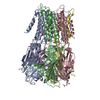
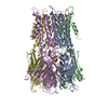
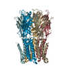
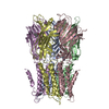
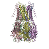
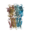
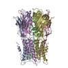
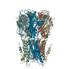
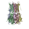
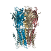
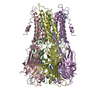
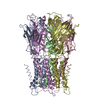
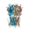
X-ray structure of a pentameric ligand gated ion channel from Erwinia chrysanthemi (ELIC) mutant L240A F247L (L9A F16L) in the presence of 10 mM cysteamine
ComponentsGamma-aminobutyric-acid receptor subunit beta-1
KeywordsTRANSPORT PROTEIN / membrane protein / liganded-gated ion channel
Function / homology
Function and homology information


extracellular ligand-gated monoatomic ion channel activity / regulation of membrane potential / transmembrane signaling receptor activity / neuron projection / signal transduction / membrane / identical protein binding
Similarity search - Function
Neurotransmitter-gated ion-channel transmembrane domain superfamily / Neuronal acetylcholine receptor / Neurotransmitter-gated ion-channel / Neurotransmitter-gated ion-channel ligand-binding domain / Neurotransmitter-gated ion-channel ligand-binding domain superfamily / Neurotransmitter-gated ion-channel ligand binding domain
Similarity search - Domain/homology
Gamma-aminobutyric-acid receptor subunit beta-1
Similarity search - Component
Biological speciesErwinia chrysanthemi (bacteria)
MethodX-RAY DIFFRACTION / SYNCHROTRON / MOLECULAR REPLACEMENT / Resolution: 4.2 Å
AuthorsGonzalez-Gutierrez, G. / Lukk, T. / Agarwal, V. / Papke, D. / Nair, S.K. / Grosman, C.
CitationJournal: Proc.Natl.Acad.Sci.USA / Year: 2012
Title: Mutations that stabilize the open state of the Erwinia chrisanthemi ligand-gated ion channel fail to change the conformation of the pore domain in crystals.
Authors: Gonzalez-Gutierrez, G. / Lukk, T. / Agarwal, V. / Papke, D. / Nair, S.K. / Grosman, C.
History
DepositionNov 19, 2011Deposition site: RCSB / Processing site: RCSB
Revision 1.0Apr 4, 2012Provider: repository / Type: Initial release
Revision 1.1Apr 18, 2012Group: Database references
Revision 1.2May 2, 2012Group: Database references
Revision 1.3Jul 17, 2019Group: Data collection / Refinement description / Category: software
Item: _software.classification / _software.contact_author ..._software.classification / _software.contact_author / _software.location / _software.name / _software.type / _software.version
Revision 1.4Sep 13, 2023Group: Data collection / Database references ...Data collection / Database references / Derived calculations / Refinement description
Category: chem_comp_atom / chem_comp_bond ...chem_comp_atom / chem_comp_bond / database_2 / pdbx_initial_refinement_model / pdbx_struct_conn_angle / struct_conn / struct_ncs_dom_lim / struct_ref_seq_dif / struct_site
Item: _database_2.pdbx_DOI / _database_2.pdbx_database_accession ..._database_2.pdbx_DOI / _database_2.pdbx_database_accession / _pdbx_struct_conn_angle.ptnr1_auth_asym_id / _pdbx_struct_conn_angle.ptnr1_label_asym_id / _pdbx_struct_conn_angle.ptnr3_auth_asym_id / _pdbx_struct_conn_angle.ptnr3_label_asym_id / _struct_conn.pdbx_dist_value / _struct_conn.ptnr1_auth_asym_id / _struct_conn.ptnr1_auth_comp_id / _struct_conn.ptnr1_auth_seq_id / _struct_conn.ptnr1_label_asym_id / _struct_conn.ptnr1_label_atom_id / _struct_conn.ptnr1_label_comp_id / _struct_conn.ptnr1_label_seq_id / _struct_conn.ptnr2_auth_asym_id / _struct_conn.ptnr2_auth_comp_id / _struct_conn.ptnr2_auth_seq_id / _struct_conn.ptnr2_label_asym_id / _struct_conn.ptnr2_label_atom_id / _struct_conn.ptnr2_label_comp_id / _struct_conn.ptnr2_label_seq_id / _struct_ncs_dom_lim.beg_auth_comp_id / _struct_ncs_dom_lim.beg_label_asym_id / _struct_ncs_dom_lim.beg_label_comp_id / _struct_ncs_dom_lim.beg_label_seq_id / _struct_ncs_dom_lim.end_auth_comp_id / _struct_ncs_dom_lim.end_label_asym_id / _struct_ncs_dom_lim.end_label_comp_id / _struct_ncs_dom_lim.end_label_seq_id / _struct_ref_seq_dif.details / _struct_site.pdbx_auth_asym_id / _struct_site.pdbx_auth_comp_id / _struct_site.pdbx_auth_seq_id

-
Structure visualization

Structure viewerMolecule:
MolmilJmol/JSmol

Downloads & links

-
Assembly

Deposited unit
A: Gamma-aminobutyric-acid receptor subunit beta-1
B: Gamma-aminobutyric-acid receptor subunit beta-1
C: Gamma-aminobutyric-acid receptor subunit beta-1
D: Gamma-aminobutyric-acid receptor subunit beta-1
E: Gamma-aminobutyric-acid receptor subunit beta-1
F: Gamma-aminobutyric-acid receptor subunit beta-1
G: Gamma-aminobutyric-acid receptor subunit beta-1
H: Gamma-aminobutyric-acid receptor subunit beta-1
I: Gamma-aminobutyric-acid receptor subunit beta-1
J: Gamma-aminobutyric-acid receptor subunit beta-1
hetero molecules


Theoretical massNumber of molelcules
Total (without water)369,49311
Polymers369,47010
Non-polymers231
Water0
1
A: Gamma-aminobutyric-acid receptor subunit beta-1
B: Gamma-aminobutyric-acid receptor subunit beta-1
C: Gamma-aminobutyric-acid receptor subunit beta-1
D: Gamma-aminobutyric-acid receptor subunit beta-1
E: Gamma-aminobutyric-acid receptor subunit beta-1


Theoretical massNumber of molelcules
Total (without water)184,7355
Polymers184,7355
Non-polymers00
Water0
TypeNameSymmetry operationNumber
identity operation1_555x,y,z1
Buried area25230 Å2
ΔGint-135 kcal/mol
Surface area65340 Å2
MethodPISA
2
F: Gamma-aminobutyric-acid receptor subunit beta-1
G: Gamma-aminobutyric-acid receptor subunit beta-1
H: Gamma-aminobutyric-acid receptor subunit beta-1
I: Gamma-aminobutyric-acid receptor subunit beta-1
J: Gamma-aminobutyric-acid receptor subunit beta-1
hetero molecules


Theoretical massNumber of molelcules
Total (without water)184,7586
Polymers184,7355
Non-polymers231
Water0
TypeNameSymmetry operationNumber
identity operation1_555x,y,z1
Buried area25360 Å2
ΔGint-148 kcal/mol
Surface area65320 Å2
MethodPISA
Unit cell
Length a, b, c (Å)105.570, 267.430, 111.160
Angle α, β, γ (deg.)90.000, 108.640, 90.000
Int Tables number4
Space group name H-MP1211
Noncrystallographic symmetry (NCS)NCS domain:
IDEns-IDDetails
11B
21C
31D
41E
51H
61A
71F
81G
91I
101J

NCS domain segments:

Component-ID: 1 / Ens-ID: 1 / Beg auth comp-ID: PRO / Beg label comp-ID: PRO / End auth comp-ID: ILE / End label comp-ID: ILE / Auth seq-ID: 11 - 317 / Label seq-ID: 13 - 319

Dom-IDSelection detailsAuth asym-IDLabel asym-ID
1chain 'B' and (resseq 11:317 )BB
2chain 'C' and (resseq 11:317 )CC
3chain 'D' and (resseq 11:317 )DD
4chain 'E' and (resseq 11:317 )EE
5chain 'H' and (resseq 11:317 )HH
6chain 'A' and (resseq 11:317 )AA
7chain 'F' and (resseq 11:317 )FF
8chain 'G' and (resseq 11:317 )GG
9chain 'I' and (resseq 11:317 )II
10chain 'J' and (resseq 11:317 )JJ

-
Components

#1: Protein
Gamma-aminobutyric-acid receptor subunit beta-1 / ELIC


Mass: 36947.035 Da / Num. of mol.: 10 / Mutation: L239A,F246L
Source method: isolated from a genetically manipulated source
Source: (gene. exp.) Erwinia chrysanthemi (bacteria) / Strain: 3937 / Gene: Dda3937_00520 / Production host: Escherichia coli (E. coli) / References: UniProt: E0SJQ4
#2: Chemical ChemComp-NA / SODIUM ION


Mass: 22.990 Da / Num. of mol.: 1 / Source method: obtained synthetically / Formula: Na

-
Experimental details

-
Experiment

ExperimentMethod: X-RAY DIFFRACTION / Number of used crystals: 1

-
Sample preparation

CrystalDensity Matthews: 4.13 Å3/Da / Density % sol: 70.22 %
Crystal growTemperature: 277 K / Method: vapor diffusion, hanging drop / pH: 6.5
Details: 10-12% PEG4000, 200 mM ammonium sulfate, 50 mM N-(2-acetamido)iminodiacetic acid (ADA), pH 6.5-6.9, VAPOR DIFFUSION, HANGING DROP, temperature 277K

-
Data collection

DiffractionMean temperature: 100 K
Diffraction sourceSource: SYNCHROTRON / Site: APS / Beamline: 21-ID-F / Wavelength: 0.97872 Å
DetectorType: MARMOSAIC 225 mm CCD / Detector: CCD / Date: Jul 1, 2011
RadiationMonochromator: Diamond(111) / Protocol: SINGLE WAVELENGTH / Monochromatic (M) / Laue (L): M / Scattering type: x-ray
Radiation wavelengthWavelength: 0.97872 Å / Relative weight: 1
ReflectionResolution: 4.2→19.988 Å / Num. obs: 42057 / % possible obs: 98.7 % / Observed criterion σ(I): -3 / Biso Wilson estimate: 103.346 Å2 / Rmerge(I) obs: 0.179 / Net I/σ(I): 16.58
Reflection shell

Diffraction-ID: 1

Resolution (Å)Highest resolution (Å)Rmerge(I) obsMean I/σ(I) obsNum. measured obsNum. unique obs% possible all
4.2-4.310.8294.1748766313999.4
4.31-4.430.6125.5148168310199.5
4.43-4.560.4797.1146743300599.9
4.56-4.70.4277.7944524286599.7
4.7-4.850.3658.9542741274999.9
4.85-5.020.3658.9441739268399.7
5.02-5.210.31910.0640366259799.7
5.21-5.420.31410.0938455247699.7
5.42-5.660.31210.337532241899.8
5.66-5.940.27511.6236167233099.7
5.94-6.260.28811.07343112214100
6.26-6.640.25512.4632061206899.5
6.64-7.10.20215.1730309196099.5
7.1-7.670.14421.1127967181599.9
7.67-8.40.08930.83258061682100
8.4-9.390.05347.2223002151399.6
9.39-10.840.03860.5920268133599.4
10.84-13.280.03367.7717106114499.5
13.28-18.780.03273.712842886100
18.780.02382.1610527715.4

-
Processing

Software
NameVersionClassificationNB
PHENIXdev_897refinement
REFMACrefinement
PDB_EXTRACT3.1data extraction
XDSdata reduction
PHASERphasing
XSCALEdata scaling
RefinementMethod to determine structure: MOLECULAR REPLACEMENT
Starting model: PDB ENTRY 2VL0
Resolution: 4.2→19.988 Å / Occupancy max: 1 / Occupancy min: 1 / SU ML: 0.48 / σ(F): 1.99 / Phase error: 22.79 / Stereochemistry target values: ML
RfactorNum. reflection% reflection
Rfree0.2494 2101 5 %
Rwork0.2198 --
obs0.2213 42030 99.8 %
Solvent computationShrinkage radii: 0.86 Å / VDW probe radii: 1.1 Å / Solvent model: FLAT BULK SOLVENT MODEL / Bsol: 84.311 Å2 / ksol: 0.246 e/Å3
Displacement parametersBiso max: 374.4 Å2 / Biso mean: 152.9963 Å2 / Biso min: 49.48 Å2
Baniso -1Baniso -2Baniso -3
1--0.0355 Å20 Å21.5061 Å2
2---0.6366 Å2-0 Å2
3---0.6721 Å2
Refinement stepCycle: LAST / Resolution: 4.2→19.988 Å
ProteinNucleic acidLigandSolventTotal
Num. atoms24990 0 1 0 24991
Refine LS restraints
Refine-IDTypeDev idealNumber
X-RAY DIFFRACTIONf_bond_d0.01225660
X-RAY DIFFRACTIONf_angle_d1.63834980
X-RAY DIFFRACTIONf_chiral_restr0.1153900
X-RAY DIFFRACTIONf_plane_restr0.0084460
X-RAY DIFFRACTIONf_dihedral_angle_d19.2989210
Refine LS restraints NCS
Ens-IDDom-IDAuth asym-IDNumberRefine-IDTypeRms dev position (Å)
11B2499X-RAY DIFFRACTIONPOSITIONAL0.055
12C2499X-RAY DIFFRACTIONPOSITIONAL0.055
13D2499X-RAY DIFFRACTIONPOSITIONAL0.053
14E2499X-RAY DIFFRACTIONPOSITIONAL0.06
15H2499X-RAY DIFFRACTIONPOSITIONAL0.052
16A2499X-RAY DIFFRACTIONPOSITIONAL0.047
17F2499X-RAY DIFFRACTIONPOSITIONAL0.054
18G2499X-RAY DIFFRACTIONPOSITIONAL0.055
19I2499X-RAY DIFFRACTIONPOSITIONAL0.054
110J2499X-RAY DIFFRACTIONPOSITIONAL0.054
LS refinement shell

Refine-ID: X-RAY DIFFRACTION / Total num. of bins used: 15

Resolution (Å)Rfactor RfreeNum. reflection RfreeRfactor RworkNum. reflection RworkNum. reflection all% reflection obs (%)
4.1999-4.29670.35571380.324426262764100
4.2967-4.4030.32631410.28826802821100
4.403-4.52070.2881390.259226352774100
4.5207-4.65220.26411400.231626762816100
4.6522-4.80030.26251410.201326612802100
4.8003-4.96940.22041380.206426372775100
4.9694-5.1650.20981400.19226572797100
5.165-5.39570.25511400.189926542794100
5.3957-5.6740.26431410.226426802821100
5.674-6.02040.32591400.257226582798100
6.0204-6.47060.30111400.256926662806100
6.4706-7.09520.28481410.224526782819100
7.0952-8.06240.17971410.164926712812100
8.0624-9.94530.15761400.151926742814100
9.9453-19.99740.25251410.23542676281799

+
About Yorodumi

-
News

-
Feb 9, 2022. New format data for meta-information of EMDB entries

New format data for meta-information of EMDB entries

  • Version 3 of the EMDB header file is now the official format.
  • The previous official version 1.9 will be removed from the archive.

Related info.:EMDB header

External links:wwPDB to switch to version 3 of the EMDB data model

-
Aug 12, 2020. Covid-19 info

Covid-19 info

URL: https://pdbj.org/emnavi/covid19.php

New page: Covid-19 featured information page in EM Navigator.

Related info.:Covid-19 info / Mar 5, 2020. Novel coronavirus structure data

+
Mar 5, 2020. Novel coronavirus structure data

Novel coronavirus structure data

Related info.:Yorodumi Speices / Aug 12, 2020. Covid-19 info

External links:COVID-19 featured content - PDBj / Molecule of the Month (242):Coronavirus Proteases

+
Jan 31, 2019. EMDB accession codes are about to change! (news from PDBe EMDB page)

EMDB accession codes are about to change! (news from PDBe EMDB page)

  • The allocation of 4 digits for EMDB accession codes will soon come to an end. Whilst these codes will remain in use, new EMDB accession codes will include an additional digit and will expand incrementally as the available range of codes is exhausted. The current 4-digit format prefixed with “EMD-” (i.e. EMD-XXXX) will advance to a 5-digit format (i.e. EMD-XXXXX), and so on. It is currently estimated that the 4-digit codes will be depleted around Spring 2019, at which point the 5-digit format will come into force.
  • The EM Navigator/Yorodumi systems omit the EMD- prefix.

Related info.:Q: What is EMD? / ID/Accession-code notation in Yorodumi/EM Navigator

External links:EMDB Accession Codes are Changing Soon! / Contact to PDBj

+
Jul 12, 2017. Major update of PDB

Major update of PDB

  • wwPDB released updated PDB data conforming to the new PDBx/mmCIF dictionary.
  • This is a major update changing the version number from 4 to 5, and with Remediation, in which all the entries are updated.
  • In this update, many items about electron microscopy experimental information are reorganized (e.g. em_software).
  • Now, EM Navigator and Yorodumi are based on the updated data.

External links:wwPDB Remediation / Enriched Model Files Conforming to OneDep Data Standards Now Available in the PDB FTP Archive

-
Yorodumi

Thousand views of thousand structures

  • Yorodumi is a browser for structure data from EMDB, PDB, SASBDB, etc.
  • This page is also the successor to EM Navigator detail page, and also detail information page/front-end page for Omokage search.
  • The word "yorodu" (or yorozu) is an old Japanese word meaning "ten thousand". "mi" (miru) is to see.

Related info.:EMDB / PDB / SASBDB / Comparison of 3 databanks / Yorodumi Search / Aug 31, 2016. New EM Navigator & Yorodumi / Yorodumi Papers / Jmol/JSmol / Function and homology information / Changes in new EM Navigator and Yorodumi

Read more