 Movie
Movie Controller

[English] 日本語
 Yorodumi
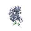
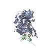
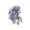
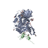
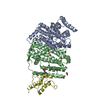
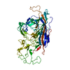
- PDB-5h9o: Complex of Murine endoplasmic reticulum alpha-glucosidase II with... -
+ Open data

- Basic information
| Entry | Database: PDB / ID: 5h9o | ||||||
|---|---|---|---|---|---|---|---|
| Title | Complex of Murine endoplasmic reticulum alpha-glucosidase II with D-Glucose | ||||||
 Components Components |
| ||||||
 Keywords Keywords |  HYDROLASE / Enzyme Glycosyl hydrolase GH31 Quality control exoglycosidase HYDROLASE / Enzyme Glycosyl hydrolase GH31 Quality control exoglycosidase | ||||||
| Function / homology |  Function and homology information Function and homology informationmannosyl-oligosaccharide alpha-1,3-glucosidase / glucan 1,3-alpha-glucosidase activity / glucosidase II complex / alpha-glucosidase activity / glucosidase activity / Regulation of Insulin-like Growth Factor (IGF) transport and uptake by Insulin-like Growth Factor Binding Proteins (IGFBPs) / Post-translational protein phosphorylation / N-glycan processing / liver development / melanosome ...mannosyl-oligosaccharide alpha-1,3-glucosidase / glucan 1,3-alpha-glucosidase activity / glucosidase II complex / alpha-glucosidase activity / glucosidase activity / Regulation of Insulin-like Growth Factor (IGF) transport and uptake by Insulin-like Growth Factor Binding Proteins (IGFBPs) / Post-translational protein phosphorylation / N-glycan processing / liver development / melanosome / negative regulation of neuron projection development / carbohydrate binding / in utero embryonic development / carbohydrate metabolic process / intracellular membrane-bounded organelle / calcium ion binding / protein-containing complex binding / Golgi apparatus / endoplasmic reticulum / RNA bindingSimilarity search - Function | ||||||
| Biological species |  Mus musculus (house mouse) Mus musculus (house mouse) | ||||||
| Method | X-RAY DIFFRACTION / SYNCHROTRON / MOLECULAR REPLACEMENT / Resolution: 2.37 Å | ||||||
 Authors Authors | Caputo, A.T. / Roversi, P. / Alonzi, D.S. / Kiappes, J.L. / Zitzmann, N. | ||||||
 Citation Citation | Journal: Proc.Natl.Acad.Sci.USA / Year: 2016 Title: Structures of mammalian ER alpha-glucosidase II capture the binding modes of broad-spectrum iminosugar antivirals. Authors: Caputo, A.T. / Alonzi, D.S. / Marti, L. / Reca, I.B. / Kiappes, J.L. / Struwe, W.B. / Cross, A. / Basu, S. / Lowe, E.D. / Darlot, B. / Santino, A. / Roversi, P. / Zitzmann, N. | ||||||
| History |
|
- Structure visualization
| Structure viewer | Molecule: MolmilJmol/JSmol |
|---|
- Downloads & links
-Download
| PDBx/mmCIF format | 5h9o.cif.gz | 725.8 KB | Display | PDBx/mmCIF format |
|---|---|---|---|---|
| PDB format | pdb5h9o.ent.gz | 611.8 KB | Display | PDB format |
| PDBx/mmJSON format | 5h9o.json.gz | Tree view | PDBx/mmJSON format | |
| Others |  Other downloads Other downloads |
-Validation report
| Arichive directory | https://data.pdbj.org/pub/pdb/validation_reports/h9/5h9oftp://data.pdbj.org/pub/pdb/validation_reports/h9/5h9o | HTTPS FTP |
|---|
-Related structure data
| Related structure data |  5f0eC 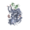 5hjoC  5hjrC  5iedC  5ieeC  5iefC 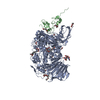 5iegC C: citing same article ( |
|---|---|
| Similar structure data |


-Links
 PDBj
PDBj


- Assembly
| Deposited unit | 
| ||||||||
|---|---|---|---|---|---|---|---|---|---|
| 1 | 
| ||||||||
| 2 | 
| ||||||||
| Unit cell |
|
-Components
-Protein , 2 types, 4 molecules ACBD
| #1: Protein | Mass: 97894.625 Da / Num. of mol.: 2 / Fragment: UNP Residues 33-944 Source method: isolated from a genetically manipulated source Source: (gene. exp.) Mus musculus (house mouse) / Gene: Ganab, G2an, Kiaa0088 / Plasmid: pHLsec / Cell line (production host): HEK293-F / Organ (production host): Kidney / Production host:  Homo sapiens (human) / References: UniProt: Q8BHN3, EC: 3.2.1.84 Homo sapiens (human) / References: UniProt: Q8BHN3, EC: 3.2.1.84#2: Protein | Glucosidases / 80K-H protein / Glucosidase II subunit beta / Protein kinase C substrate 60.1 kDa protein heavy chain / PKCSHMass: 9568.298 Da / Num. of mol.: 2 / Fragment: UNP residues 30-117 Source method: isolated from a genetically manipulated source Source: (gene. exp.) Mus musculus (house mouse) / Gene: Prkcsh / Plasmid: pHLsec / Cell line (production host): HEK293-F / Production host: Homo sapiens (human) / References: UniProt: O08795 |
|---|
-Sugars , 2 types, 6 molecules 

| #3: Sugar | ChemComp-NAG / N-Acetylglucosamine Type: D-saccharide, beta linking / Mass: 221.208 Da / Num. of mol.: 4 Type: D-saccharide, beta linking / Mass: 221.208 Da / Num. of mol.: 4Source method: isolated from a genetically manipulated source Formula: C8H15NO6 #9: Sugar | Glucose Type: D-saccharide, alpha linking / Mass: 180.156 Da / Num. of mol.: 2 Type: D-saccharide, alpha linking / Mass: 180.156 Da / Num. of mol.: 2Source method: isolated from a genetically manipulated source Formula: C6H12O6 |
|---|
-Non-polymers , 8 types, 476 molecules 


| #4: Chemical | ChemComp-EDO / Ethylene glycol Mass: 62.068 Da / Num. of mol.: 8 Mass: 62.068 Da / Num. of mol.: 8Source method: isolated from a genetically manipulated source Formula: C2H6O2 #5: Chemical | ChemComp-FMT / Formic acid Mass: 46.025 Da / Num. of mol.: 19 Mass: 46.025 Da / Num. of mol.: 19Source method: isolated from a genetically manipulated source Formula: CH2O2 #6: Chemical | ChemComp-PG4 / Polyethylene glycol Mass: 194.226 Da / Num. of mol.: 7 / Source method: obtained synthetically / Formula: C8H18O5 / Comment: precipitant*YM Mass: 194.226 Da / Num. of mol.: 7 / Source method: obtained synthetically / Formula: C8H18O5 / Comment: precipitant*YM#7: Chemical | ChemComp-P6G / Polyethylene glycol Mass: 282.331 Da / Num. of mol.: 5 / Source method: obtained synthetically / Formula: C12H26O7 / Comment: precipitant*YM Mass: 282.331 Da / Num. of mol.: 5 / Source method: obtained synthetically / Formula: C12H26O7 / Comment: precipitant*YM#8: Chemical | ChemComp-OXM / | Oxamic acid Mass: 89.050 Da / Num. of mol.: 1 / Source method: obtained synthetically / Formula: C2H3NO3 Mass: 89.050 Da / Num. of mol.: 1 / Source method: obtained synthetically / Formula: C2H3NO3#10: Chemical | ChemComp-CA /  Mass: 40.078 Da / Num. of mol.: 4 / Source method: obtained synthetically / Formula: Ca Mass: 40.078 Da / Num. of mol.: 4 / Source method: obtained synthetically / Formula: Ca#11: Chemical | ChemComp-TAR / | Tartaric acid Mass: 150.087 Da / Num. of mol.: 1 / Source method: obtained synthetically / Formula: C4H6O6 Mass: 150.087 Da / Num. of mol.: 1 / Source method: obtained synthetically / Formula: C4H6O6#12: Water | ChemComp-HOH / | WaterMass: 18.015 Da / Num. of mol.: 431 / Source method: isolated from a natural source / Formula: H2O |
|---|
-Experimental details
-Experiment
| Experiment | Method: X-RAY DIFFRACTION / Number of used crystals: 1 |
|---|
- Sample preparation
| Crystal | Density Matthews: 2.3 Å3/Da / Density % sol: 46.61 % |
|---|---|
| Crystal grow | Temperature: 291 K / Method: vapor diffusion, sitting drop / pH: 6.25 Details: 32 % Morpheus ethylene glycol/PEG 8000 mix, 0.05 M Morpheus carboxylic acids mix, 0.1 M Morpheus buffer system 1 |
-Data collection
| Diffraction | Mean temperature: 100 K |
|---|---|
| Diffraction source | Source: SYNCHROTRON / Site: Diamond  / Beamline: I04 / Wavelength: 0.9795 Å / Beamline: I04 / Wavelength: 0.9795 Å |
| Detector | Type: DECTRIS PILATUS 6M-F / Detector: PIXEL / Date: Sep 27, 2015 |
| Radiation | Protocol: SINGLE WAVELENGTH / Monochromatic (M) / Laue (L): M / Scattering type: x-ray |
| Radiation wavelength | Wavelength: 0.9795 Å / Relative weight: 1 |
| Reflection | Resolution: 2.37→87.88 Å / Num. obs: 89708 / % possible obs: 99.8 % / Redundancy: 5.5 % / Biso Wilson estimate: 33.37 Å2 / Rsym value: 0.24 / Net I/σ(I): 5.1 |
| Reflection shell | Resolution: 2.37→2.43 Å / Redundancy: 5.4 % / Mean I/σ(I) obs: 1.2 / Num. unique all: 6578 / Rsym value: 1.53 / % possible all: 99.7 |
- Processing
| Software |
| ||||||||||||||||||||||||||||||||||||||||||||||||||||||||||||||||||||||||||||||||||||||||||||||||||||||||||||||||||
|---|---|---|---|---|---|---|---|---|---|---|---|---|---|---|---|---|---|---|---|---|---|---|---|---|---|---|---|---|---|---|---|---|---|---|---|---|---|---|---|---|---|---|---|---|---|---|---|---|---|---|---|---|---|---|---|---|---|---|---|---|---|---|---|---|---|---|---|---|---|---|---|---|---|---|---|---|---|---|---|---|---|---|---|---|---|---|---|---|---|---|---|---|---|---|---|---|---|---|---|---|---|---|---|---|---|---|---|---|---|---|---|---|---|---|---|
| Refinement | Method to determine structure: MOLECULAR REPLACEMENT / Resolution: 2.37→87.66 Å / Cor.coef. Fo:Fc: 0.905 / Cor.coef. Fo:Fc free: 0.882 / Rfactor Rfree error: 0 / Cross valid method: THROUGHOUT / σ(F): 0 / SU R Blow DPI: 0.414 / SU Rfree Blow DPI: 0.25
| ||||||||||||||||||||||||||||||||||||||||||||||||||||||||||||||||||||||||||||||||||||||||||||||||||||||||||||||||||
| Displacement parameters | Biso mean: 33.8 Å2
| ||||||||||||||||||||||||||||||||||||||||||||||||||||||||||||||||||||||||||||||||||||||||||||||||||||||||||||||||||
| Refine analyze | Luzzati coordinate error obs: 0.37 Å | ||||||||||||||||||||||||||||||||||||||||||||||||||||||||||||||||||||||||||||||||||||||||||||||||||||||||||||||||||
| Refinement step | Cycle: LAST / Resolution: 2.37→87.66 Å
| ||||||||||||||||||||||||||||||||||||||||||||||||||||||||||||||||||||||||||||||||||||||||||||||||||||||||||||||||||
| Refine LS restraints |
| ||||||||||||||||||||||||||||||||||||||||||||||||||||||||||||||||||||||||||||||||||||||||||||||||||||||||||||||||||
| LS refinement shell | Resolution: 2.37→2.43 Å / Rfactor Rfree error: 0 / Total num. of bins used: 20
|
