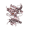
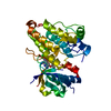
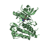
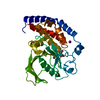
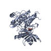
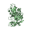
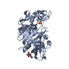
[English] 日本語
Yorodumi
- PDB-5h3q: Crystal Structure of TrkA kinase with ligand -

+
Open data


ID or keywords:

Loading...

-
Basic information

Entry
Database: PDB / ID: 5h3q
TitleCrystal Structure of TrkA kinase with ligand
ComponentsHigh affinity nerve growth factor receptor
KeywordsTRANSFERASE/TRANSFERASE INHIBITOR / TrkA inhibitor / TRANSFERASE-TRANSFERASE INHIBITOR complex
Function / homology
Function and homology information


behavioral response to formalin induced pain / neurotrophin p75 receptor binding / olfactory nerve development / response to hydrostatic pressure / TRKA activation by NGF / PLC-gamma1 signalling / programmed cell death involved in cell development / Signalling to STAT3 / neurotrophin receptor activity / mechanoreceptor differentiation ...behavioral response to formalin induced pain / neurotrophin p75 receptor binding / olfactory nerve development / response to hydrostatic pressure / TRKA activation by NGF / PLC-gamma1 signalling / programmed cell death involved in cell development / Signalling to STAT3 / neurotrophin receptor activity / mechanoreceptor differentiation / nerve growth factor receptor activity / neurotrophin binding / Sertoli cell development / axonogenesis involved in innervation / GPI-linked ephrin receptor activity / nerve growth factor signaling pathway / Retrograde neurotrophin signalling / nerve growth factor binding / NGF-independant TRKA activation / sympathetic nervous system development / Signalling to p38 via RIT and RIN / ARMS-mediated activation / positive regulation of Ras protein signal transduction / positive regulation of programmed cell death / positive regulation of synapse assembly / PI3K/AKT activation / Frs2-mediated activation / regulation of phosphatidylinositol 3-kinase/protein kinase B signal transduction / detection of temperature stimulus involved in sensory perception of pain / neurotrophin TRK receptor signaling pathway / response to axon injury / neuron development / Signalling to RAS / detection of mechanical stimulus involved in sensory perception of pain / response to electrical stimulus / peptidyl-tyrosine autophosphorylation / transmembrane receptor protein tyrosine kinase activity / positive regulation of synaptic transmission, glutamatergic / response to nutrient levels / B cell differentiation / cellular response to nerve growth factor stimulus / axon guidance / receptor protein-tyrosine kinase / positive regulation of GTPase activity / kinase binding / positive regulation of neuron projection development / cellular response to nicotine / circadian rhythm / peptidyl-tyrosine phosphorylation / recycling endosome membrane / positive regulation of angiogenesis / neuron projection development / late endosome / late endosome membrane / positive regulation of NF-kappaB transcription factor activity / early endosome membrane / protein tyrosine kinase activity / neuron apoptotic process / negative regulation of neuron apoptotic process / protein autophosphorylation / early endosome / learning or memory / positive regulation of ERK1 and ERK2 cascade / receptor complex / endosome membrane / response to xenobiotic stimulus / positive regulation of protein phosphorylation / negative regulation of cell population proliferation / axon / protein phosphorylation / neuronal cell body / dendrite / negative regulation of apoptotic process / cell surface / protein homodimerization activity / protein-containing complex / ATP binding / identical protein binding / plasma membrane
Similarity search - Function
High affinity nerve growth factor receptor NTRK1 / Tyrosine kinase receptor A, transmembrane domain / Tyrosine kinase receptor A trans-membrane domain / Growth factor receptor NTRK / Growth factor receptor NTRK, leucine rich repeat C-terminal / Leucine rich repeat C-terminal motif / Cysteine-rich flanking region, C-terminal / Leucine rich repeat C-terminal domain / Tyrosine-protein kinase, receptor class II, conserved site / Receptor tyrosine kinase class II signature. ...High affinity nerve growth factor receptor NTRK1 / Tyrosine kinase receptor A, transmembrane domain / Tyrosine kinase receptor A trans-membrane domain / Growth factor receptor NTRK / Growth factor receptor NTRK, leucine rich repeat C-terminal / Leucine rich repeat C-terminal motif / Cysteine-rich flanking region, C-terminal / Leucine rich repeat C-terminal domain / Tyrosine-protein kinase, receptor class II, conserved site / Receptor tyrosine kinase class II signature. / Leucine rich repeat / Leucine-rich repeat / Leucine-rich repeat domain superfamily / Tyrosine-protein kinase, catalytic domain / Tyrosine kinase, catalytic domain / Tyrosine protein kinases specific active-site signature. / Tyrosine-protein kinase, active site / Protein tyrosine and serine/threonine kinase / Serine-threonine/tyrosine-protein kinase, catalytic domain / Transferase(Phosphotransferase) domain 1 / Transferase(Phosphotransferase); domain 1 / Phosphorylase Kinase; domain 1 / Phosphorylase Kinase; domain 1 / Ig-like domain profile. / Immunoglobulin-like domain / Immunoglobulin-like domain superfamily / Protein kinase, ATP binding site / Protein kinases ATP-binding region signature. / Immunoglobulin-like fold / Protein kinase domain profile. / Protein kinase domain / Protein kinase-like domain superfamily / 2-Layer Sandwich / Orthogonal Bundle / Mainly Alpha / Alpha Beta
Similarity search - Domain/homology
Chem-7HF / 2,3-DIHYDROXY-1,4-DITHIOBUTANE / High affinity nerve growth factor receptor
Similarity search - Component
Biological speciesHomo sapiens (human)
MethodX-RAY DIFFRACTION / SYNCHROTRON / MOLECULAR REPLACEMENT / Resolution: 2.1 Å
AuthorsNoritaka, F.
CitationJournal: Bioorg. Med. Chem. Lett. / Year: 2017
Title: The juxtamembrane region of TrkA kinase is critical for inhibitor selectivity
Authors: Furuya, N. / Momose, T. / Katsuno, K. / Fushimi, N. / Muranaka, H. / Handa, C. / Ozawa, T. / Kinoshita, T.
History
DepositionOct 26, 2016Deposition site: PDBJ / Processing site: PDBJ
Revision 1.0Feb 22, 2017Provider: repository / Type: Initial release
Revision 1.1Mar 8, 2017Group: Database references
Revision 1.2Feb 26, 2020Group: Data collection / Category: diffrn_source / Item: _diffrn_source.pdbx_synchrotron_site
Revision 1.3Nov 8, 2023Group: Data collection / Database references ...Data collection / Database references / Derived calculations / Refinement description
Category: chem_comp_atom / chem_comp_bond ...chem_comp_atom / chem_comp_bond / database_2 / pdbx_initial_refinement_model / struct_conn
Item: _database_2.pdbx_DOI / _database_2.pdbx_database_accession ..._database_2.pdbx_DOI / _database_2.pdbx_database_accession / _struct_conn.conn_type_id / _struct_conn.id / _struct_conn.pdbx_dist_value / _struct_conn.pdbx_leaving_atom_flag / _struct_conn.ptnr1_auth_comp_id / _struct_conn.ptnr1_auth_seq_id / _struct_conn.ptnr1_label_atom_id / _struct_conn.ptnr1_label_comp_id / _struct_conn.ptnr1_label_seq_id / _struct_conn.ptnr2_auth_comp_id / _struct_conn.ptnr2_auth_seq_id / _struct_conn.ptnr2_label_asym_id / _struct_conn.ptnr2_label_atom_id / _struct_conn.ptnr2_label_comp_id

-
Structure visualization

Structure viewerMolecule:
MolmilJmol/JSmol

Downloads & links

-
Assembly

Deposited unit
A: High affinity nerve growth factor receptor
hetero molecules


Theoretical massNumber of molelcules
Total (without water)37,3265
Polymers36,4951
Non-polymers8314
Water1,69394
1


  • Idetical with deposited unit
  • defined by author&software
TypeNameSymmetry operationNumber
identity operation1_555x,y,z1
Buried area140 Å2
ΔGint-12 kcal/mol
Surface area14550 Å2
MethodPISA
Unit cell
Length a, b, c (Å)140.845, 157.195, 52.306
Angle α, β, γ (deg.)90.000, 90.000, 90.000
Int Tables number21
Space group name H-MC222

-
Components

#1: Protein High affinity nerve growth factor receptor / TrkA kinase / Neurotrophic tyrosine kinase receptor type 1 / TRK1-transforming tyrosine kinase ...TrkA kinase / Neurotrophic tyrosine kinase receptor type 1 / TRK1-transforming tyrosine kinase protein / Tropomyosin-related kinase A / Tyrosine kinase receptor / Tyrosine kinase receptor A / Trk-A / gp140trk / p140-TrkA


Mass: 36494.926 Da / Num. of mol.: 1
Source method: isolated from a genetically manipulated source
Source: (gene. exp.) Homo sapiens (human) / Gene: NTRK1, MTC, TRK, TRKA / Production host: Trichoplusia ni (cabbage looper)
References: UniProt: P04629, receptor protein-tyrosine kinase
#2: Chemical ChemComp-7HF / 1-[(3S,4R)-4-[3,4-bis(fluoranyl)phenyl]-1-(2-methoxyethyl)pyrrolidin-3-yl]-3-(5-ethoxy-4-methyl-2-phenyl-pyrazol-3-yl)urea


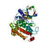
Mass: 499.553 Da / Num. of mol.: 1 / Source method: obtained synthetically / Formula: C26H31F2N5O3
#3: Chemical ChemComp-DTT / 2,3-DIHYDROXY-1,4-DITHIOBUTANE / 1,4-DITHIOTHREITOL / Dithiothreitol


Mass: 154.251 Da / Num. of mol.: 2 / Source method: obtained synthetically / Formula: C4H10O2S2
#4: Chemical ChemComp-NA / SODIUM ION


Mass: 22.990 Da / Num. of mol.: 1 / Source method: obtained synthetically / Formula: Na
#5: Water ChemComp-HOH / water / Water


Mass: 18.015 Da / Num. of mol.: 94 / Source method: isolated from a natural source / Formula: H2O

-
Experimental details

-
Experiment

ExperimentMethod: X-RAY DIFFRACTION / Number of used crystals: 1

-
Sample preparation

CrystalDensity Matthews: 3.97 Å3/Da / Density % sol: 68.99 %
Crystal growTemperature: 293 K / Method: vapor diffusion, sitting drop / pH: 8.5
Details: 1.2M Sodium Tartrate, 100mM Tris-HCl pH 8.5, 5 mM DTT

-
Data collection

DiffractionMean temperature: 100 K
Diffraction sourceSource: SYNCHROTRON / Site: SPring-8 / Beamline: BL41XU / Wavelength: 1 Å
DetectorType: DECTRIS PILATUS 6M / Detector: PIXEL / Date: Jul 15, 2014
RadiationProtocol: SINGLE WAVELENGTH / Monochromatic (M) / Laue (L): M / Scattering type: x-ray
Radiation wavelengthWavelength: 1 Å / Relative weight: 1
ReflectionResolution: 2.1→49.1 Å / Num. obs: 32612 / % possible obs: 99.67 % / Redundancy: 6.6 % / Rmerge(I) obs: 0.114 / Net I/σ(I): 13.98
Reflection shellResolution: 2.1→2.16 Å / Rmerge(I) obs: 1.688 / Rejects: 0

-
Processing

Software
NameVersionClassification
REFMAC5.8.0155refinement
XDSdata reduction
XDSdata scaling
MOLREPphasing
RefinementMethod to determine structure: MOLECULAR REPLACEMENT
Starting model: 4F0I
Resolution: 2.1→46.81 Å / Cor.coef. Fo:Fc: 0.958 / Cor.coef. Fo:Fc free: 0.953 / WRfactor Rfree: 0.1909 / WRfactor Rwork: 0.1665 / FOM work R set: 0.7829 / SU B: 4.599 / SU ML: 0.112 / SU R Cruickshank DPI: 0.137 / SU Rfree: 0.1295 / Cross valid method: THROUGHOUT / σ(F): 0 / ESU R: 0.137 / ESU R Free: 0.129 / Stereochemistry target values: MAXIMUM LIKELIHOOD
Details: HYDROGENS HAVE BEEN ADDED IN THE RIDING POSITIONS U VALUES : REFINED INDIVIDUALLY
RfactorNum. reflection% reflectionSelection details
Rfree0.2151 1704 5 %RANDOM
Rwork0.1884 ---
obs0.1898 32367 99.48 %-
Solvent computationIon probe radii: 0.8 Å / Shrinkage radii: 0.8 Å / VDW probe radii: 1.2 Å / Solvent model: MASK
Displacement parametersBiso max: 120.09 Å2 / Biso mean: 45.184 Å2 / Biso min: 23.4 Å2
Baniso -1Baniso -2Baniso -3
1-2.93 Å20 Å2-0 Å2
2---0.98 Å20 Å2
3----1.95 Å2
Refinement stepCycle: final / Resolution: 2.1→46.81 Å
ProteinNucleic acidLigandSolventTotal
Num. atoms2387 0 53 94 2534
Biso mean--49.85 47.57 -
Num. residues----303
Refine LS restraints
Refine-IDTypeDev idealDev ideal targetNumber
X-RAY DIFFRACTIONr_bond_refined_d0.0220.0192518
X-RAY DIFFRACTIONr_bond_other_d0.0040.022382
X-RAY DIFFRACTIONr_angle_refined_deg2.1371.9683412
X-RAY DIFFRACTIONr_angle_other_deg1.2873.0065449
X-RAY DIFFRACTIONr_dihedral_angle_1_deg6.6285303
X-RAY DIFFRACTIONr_dihedral_angle_2_deg36.23322.931116
X-RAY DIFFRACTIONr_dihedral_angle_3_deg15.44115403
X-RAY DIFFRACTIONr_dihedral_angle_4_deg15.0331520
X-RAY DIFFRACTIONr_chiral_restr0.1330.2364
X-RAY DIFFRACTIONr_gen_planes_refined0.0120.0212838
X-RAY DIFFRACTIONr_gen_planes_other0.0020.02615
X-RAY DIFFRACTIONr_mcbond_it3.9434.1471216
X-RAY DIFFRACTIONr_mcbond_other3.9424.1491217
X-RAY DIFFRACTIONr_mcangle_it5.5616.1911519
LS refinement shellResolution: 2.104→2.159 Å / Total num. of bins used: 20
RfactorNum. reflection% reflection
Rfree0.424 120 -
Rwork0.385 2285 -
all-2405 -
obs--97.13 %

+
About Yorodumi

-
News

-
Feb 9, 2022. New format data for meta-information of EMDB entries

New format data for meta-information of EMDB entries

  • Version 3 of the EMDB header file is now the official format.
  • The previous official version 1.9 will be removed from the archive.

Related info.:EMDB header

External links:wwPDB to switch to version 3 of the EMDB data model

-
Aug 12, 2020. Covid-19 info

Covid-19 info

URL: https://pdbj.org/emnavi/covid19.php

New page: Covid-19 featured information page in EM Navigator.

Related info.:Covid-19 info / Mar 5, 2020. Novel coronavirus structure data

+
Mar 5, 2020. Novel coronavirus structure data

Novel coronavirus structure data

Related info.:Yorodumi Speices / Aug 12, 2020. Covid-19 info

External links:COVID-19 featured content - PDBj / Molecule of the Month (242):Coronavirus Proteases

+
Jan 31, 2019. EMDB accession codes are about to change! (news from PDBe EMDB page)

EMDB accession codes are about to change! (news from PDBe EMDB page)

  • The allocation of 4 digits for EMDB accession codes will soon come to an end. Whilst these codes will remain in use, new EMDB accession codes will include an additional digit and will expand incrementally as the available range of codes is exhausted. The current 4-digit format prefixed with “EMD-” (i.e. EMD-XXXX) will advance to a 5-digit format (i.e. EMD-XXXXX), and so on. It is currently estimated that the 4-digit codes will be depleted around Spring 2019, at which point the 5-digit format will come into force.
  • The EM Navigator/Yorodumi systems omit the EMD- prefix.

Related info.:Q: What is EMD? / ID/Accession-code notation in Yorodumi/EM Navigator

External links:EMDB Accession Codes are Changing Soon! / Contact to PDBj

+
Jul 12, 2017. Major update of PDB

Major update of PDB

  • wwPDB released updated PDB data conforming to the new PDBx/mmCIF dictionary.
  • This is a major update changing the version number from 4 to 5, and with Remediation, in which all the entries are updated.
  • In this update, many items about electron microscopy experimental information are reorganized (e.g. em_software).
  • Now, EM Navigator and Yorodumi are based on the updated data.

External links:wwPDB Remediation / Enriched Model Files Conforming to OneDep Data Standards Now Available in the PDB FTP Archive

-
Yorodumi

Thousand views of thousand structures

  • Yorodumi is a browser for structure data from EMDB, PDB, SASBDB, etc.
  • This page is also the successor to EM Navigator detail page, and also detail information page/front-end page for Omokage search.
  • The word "yorodu" (or yorozu) is an old Japanese word meaning "ten thousand". "mi" (miru) is to see.

Related info.:EMDB / PDB / SASBDB / Comparison of 3 databanks / Yorodumi Search / Aug 31, 2016. New EM Navigator & Yorodumi / Yorodumi Papers / Jmol/JSmol / Function and homology information / Changes in new EM Navigator and Yorodumi

Read more