 Movie
Movie Controller

[English] 日本語
 Yorodumi
- PDB-5gv0: Crystal structure of the membrane-proximal domain of mouse lysoso... -
+ Open data

- Basic information
| Entry | Database: PDB / ID: 5gv0 | ||||||
|---|---|---|---|---|---|---|---|
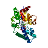
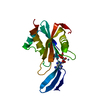
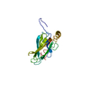
| Title | Crystal structure of the membrane-proximal domain of mouse lysosome-associated membrane protein 1 (LAMP-1) | ||||||
 Components Components | Lysosome-associated membrane glycoprotein 1 | ||||||
 Keywords Keywords | MEMBRANE PROTEIN | ||||||
| Function / homology |  Function and homology information Function and homology informationregulation of organelle transport along microtubule / : / granzyme-mediated programmed cell death signaling pathway / phagolysosome membrane / autophagic cell death / alveolar lamellar body / cytolytic granule membrane / cytolytic granule / positive regulation of natural killer cell degranulation / establishment of protein localization to organelle ...regulation of organelle transport along microtubule / : / granzyme-mediated programmed cell death signaling pathway / phagolysosome membrane / autophagic cell death / alveolar lamellar body / cytolytic granule membrane / cytolytic granule / positive regulation of natural killer cell degranulation / establishment of protein localization to organelle / Golgi to lysosome transport / vacuole / autolysosome / positive regulation of natural killer cell mediated cytotoxicity / autophagosome membrane / phagocytic vesicle / Neutrophil degranulation / multivesicular body / sarcolemma / melanosome / synaptic vesicle / late endosome / late endosome membrane / cytoplasmic vesicle / spermatogenesis / vesicle / lysosome / protein stabilization / endosome membrane / endosome / lysosomal membrane / protein domain specific binding / external side of plasma membrane / neuronal cell body / dendrite / perinuclear region of cytoplasm / enzyme binding / cell surface / plasma membrane / cytosol / cytoplasmSimilarity search - Function | ||||||
| Biological species |  Mus musculus (house mouse) Mus musculus (house mouse) | ||||||
| Method | X-RAY DIFFRACTION / SYNCHROTRON / SAD / Resolution: 1.5 Å | ||||||
 Authors Authors | Tomabechi, Y. / Ehara, H. / Kukimoto-Niino, M. / Shirouzu, M. | ||||||
 Citation Citation | Journal: Biochem.Biophys.Res.Commun. / Year: 2016 Title: Lysosome-associated membrane proteins-1 and -2 (LAMP-1 and LAMP-2) assemble via distinct modes Authors: Terasawa, K. / Tomabechi, Y. / Ikeda, M. / Ehara, H. / Kukimoto-Niino, M. / Wakiyama, M. / Podyma-Inoue, K.A. / Rajapakshe, A.R. / Watabe, T. / Shirouzu, M. / Hara-Yokoyama, M. | ||||||
| History |
|
- Structure visualization
| Structure viewer | Molecule: MolmilJmol/JSmol |
|---|
- Downloads & links
-Download
| PDBx/mmCIF format | 5gv0.cif.gz | 48.9 KB | Display | PDBx/mmCIF format |
|---|---|---|---|---|
| PDB format | pdb5gv0.ent.gz | 35.9 KB | Display | PDB format |
| PDBx/mmJSON format | 5gv0.json.gz | Tree view | PDBx/mmJSON format | |
| Others |  Other downloads Other downloads |
-Validation report
| Arichive directory | https://data.pdbj.org/pub/pdb/validation_reports/gv/5gv0ftp://data.pdbj.org/pub/pdb/validation_reports/gv/5gv0 | HTTPS FTP |
|---|
-Related structure data


-Links
 PDBj
PDBj


- Assembly
| Deposited unit | 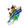
| ||||||||
|---|---|---|---|---|---|---|---|---|---|
| 1 |
| ||||||||
| Unit cell |
|
-Components
| #1: Protein | / Lysosome-associated membrane protein 1 / 120 kDa lysosomal membrane glycoprotein / CD107 antigen- ...Lysosome-associated membrane protein 1 / 120 kDa lysosomal membrane glycoprotein / CD107 antigen-like family member A / LGP-120 / Lysosomal membrane glycoprotein A / LGP-A / P2B Mass: 18253.611 Da / Num. of mol.: 1 / Fragment: UNP residues 208-370 Source method: isolated from a genetically manipulated source Source: (gene. exp.) Mus musculus (house mouse) / Gene: Lamp1, Lamp-1 / Plasmid: pOriP / Cell line (production host): HEK293 / Production host:  Homo sapiens (human) / References: UniProt: P11438 Homo sapiens (human) / References: UniProt: P11438 | ||||
|---|---|---|---|---|---|
| #2: Chemical | Sulfate  Mass: 96.063 Da / Num. of mol.: 2 Mass: 96.063 Da / Num. of mol.: 2Source method: isolated from a genetically manipulated source Formula: SO4 #3: Sugar | ChemComp-NAG / N-Acetylglucosamine  Type: D-saccharide, beta linking / Mass: 221.208 Da / Num. of mol.: 4 / Source method: obtained synthetically / Formula: C8H15NO6 Type: D-saccharide, beta linking / Mass: 221.208 Da / Num. of mol.: 4 / Source method: obtained synthetically / Formula: C8H15NO6#4: Water | ChemComp-HOH / | Water Mass: 18.015 Da / Num. of mol.: 201 / Source method: isolated from a natural source / Formula: H2O Mass: 18.015 Da / Num. of mol.: 201 / Source method: isolated from a natural source / Formula: H2O |
-Experimental details
-Experiment
| Experiment | Method: X-RAY DIFFRACTION / Number of used crystals: 1 |
|---|
- Sample preparation
| Crystal | Density Matthews: 2.13 Å3/Da / Density % sol: 42.18 % |
|---|---|
| Crystal grow | Temperature: 293 K / Method: vapor diffusion, sitting drop Details: 2M ammonium sulphate, 0.1M sodium HEPES (pH 7.5), 2% (v/v) polyethylene glycol 400 |
-Data collection
| Diffraction | Mean temperature: 100 K | ||||||||||||||||||||||||||||||||||||||||||||||||||||||||||||||||||||||||||||||||||||||||||||||||||||||||||||||||||||||||||||||
|---|---|---|---|---|---|---|---|---|---|---|---|---|---|---|---|---|---|---|---|---|---|---|---|---|---|---|---|---|---|---|---|---|---|---|---|---|---|---|---|---|---|---|---|---|---|---|---|---|---|---|---|---|---|---|---|---|---|---|---|---|---|---|---|---|---|---|---|---|---|---|---|---|---|---|---|---|---|---|---|---|---|---|---|---|---|---|---|---|---|---|---|---|---|---|---|---|---|---|---|---|---|---|---|---|---|---|---|---|---|---|---|---|---|---|---|---|---|---|---|---|---|---|---|---|---|---|---|
| Diffraction source | Source: SYNCHROTRON / Site: SPring-8  / Beamline: BL26B2 / Wavelength: 1 Å / Beamline: BL26B2 / Wavelength: 1 Å | ||||||||||||||||||||||||||||||||||||||||||||||||||||||||||||||||||||||||||||||||||||||||||||||||||||||||||||||||||||||||||||||
| Detector | Type: MARMOSAIC 225 mm CCD / Detector: CCD / Date: Oct 6, 2014 | ||||||||||||||||||||||||||||||||||||||||||||||||||||||||||||||||||||||||||||||||||||||||||||||||||||||||||||||||||||||||||||||
| Radiation | Protocol: SINGLE WAVELENGTH / Monochromatic (M) / Laue (L): M / Scattering type: x-ray | ||||||||||||||||||||||||||||||||||||||||||||||||||||||||||||||||||||||||||||||||||||||||||||||||||||||||||||||||||||||||||||||
| Radiation wavelength | Wavelength: 1 Å / Relative weight: 1 | ||||||||||||||||||||||||||||||||||||||||||||||||||||||||||||||||||||||||||||||||||||||||||||||||||||||||||||||||||||||||||||||
| Reflection | Resolution: 1.5→50 Å / Num. obs: 24809 / % possible obs: 99.3 % / Redundancy: 6.6 % / Rmerge(I) obs: 0.059 / Net I/av σ(I): 50.639 / Net I/σ(I): 16.4 | ||||||||||||||||||||||||||||||||||||||||||||||||||||||||||||||||||||||||||||||||||||||||||||||||||||||||||||||||||||||||||||||
| Reflection shell |
|
- Processing
| Software |
| |||||||||||||||||||||||||||||||||||||||||||||||||||||||||||||||||||||||||||||||||||||||||||||||||||||||||
|---|---|---|---|---|---|---|---|---|---|---|---|---|---|---|---|---|---|---|---|---|---|---|---|---|---|---|---|---|---|---|---|---|---|---|---|---|---|---|---|---|---|---|---|---|---|---|---|---|---|---|---|---|---|---|---|---|---|---|---|---|---|---|---|---|---|---|---|---|---|---|---|---|---|---|---|---|---|---|---|---|---|---|---|---|---|---|---|---|---|---|---|---|---|---|---|---|---|---|---|---|---|---|---|---|---|---|
| Refinement | Method to determine structure: SAD / Resolution: 1.5→34.48 Å / SU ML: 0.12 / Cross valid method: FREE R-VALUE / σ(F): 1.39 / Phase error: 18.62
| |||||||||||||||||||||||||||||||||||||||||||||||||||||||||||||||||||||||||||||||||||||||||||||||||||||||||
| Solvent computation | Shrinkage radii: 0.9 Å / VDW probe radii: 1.11 Å | |||||||||||||||||||||||||||||||||||||||||||||||||||||||||||||||||||||||||||||||||||||||||||||||||||||||||
| Displacement parameters | Biso max: 79.35 Å2 / Biso mean: 19.8439 Å2 / Biso min: 7.86 Å2 | |||||||||||||||||||||||||||||||||||||||||||||||||||||||||||||||||||||||||||||||||||||||||||||||||||||||||
| Refinement step | Cycle: final / Resolution: 1.5→34.48 Å
| |||||||||||||||||||||||||||||||||||||||||||||||||||||||||||||||||||||||||||||||||||||||||||||||||||||||||
| Refine LS restraints |
| |||||||||||||||||||||||||||||||||||||||||||||||||||||||||||||||||||||||||||||||||||||||||||||||||||||||||
| LS refinement shell | Refine-ID: X-RAY DIFFRACTION / Total num. of bins used: 14
|
