 Movie
Movie Controller


+ Open data

- Basic information
| Entry | Database: PDB / ID: 5gox | ||||||
|---|---|---|---|---|---|---|---|
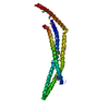
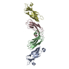
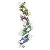
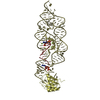
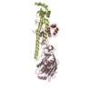
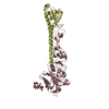
| Title | Eukaryotic Rad50 Functions as A Rod-shaped Dimer | ||||||
 Components Components | DNA repair protein RAD50 | ||||||
 Keywords Keywords | HYDROLASE / DNA repair | ||||||
| Function / homology |  Function and homology informationchromosome, telomeric region => GO:0000781 / telomeric 3' overhang formation / : / Mre11 complex / negative regulation of telomere capping / Sensing of DNA Double Strand Breaks / regulation of mitotic recombination / chromosome organization involved in meiotic cell cycle / Hydrolases; Acting on acid anhydrides / chromatin => GO:0000785 ...chromosome, telomeric region => GO:0000781 / telomeric 3' overhang formation / : / Mre11 complex / negative regulation of telomere capping / Sensing of DNA Double Strand Breaks / regulation of mitotic recombination / chromosome organization involved in meiotic cell cycle / Hydrolases; Acting on acid anhydrides / chromatin => GO:0000785 / G-quadruplex DNA binding / adenylate kinase activity / double-stranded telomeric DNA binding / DNA double-strand break processing / telomere maintenance via recombination / positive regulation of telomere maintenance / single-stranded telomeric DNA binding / HDR through MMEJ (alt-NHEJ) / telomere capping / positive regulation of kinase activity / reciprocal meiotic recombination / Homologous DNA Pairing and Strand Exchange / Resolution of D-loop Structures through Synthesis-Dependent Strand Annealing (SDSA) / Resolution of D-loop Structures through Holliday Junction Intermediates / DNA duplex unwinding / HDR through Single Strand Annealing (SSA) / Presynaptic phase of homologous DNA pairing and strand exchange / telomere maintenance via telomerase / positive regulation of protein autophosphorylation / viral process / telomere maintenance / regulation of signal transduction by p53 class mediator / condensed nuclear chromosome / Nonhomologous End-Joining (NHEJ) / double-strand break repair via homologous recombination / HDR through Homologous Recombination (HRR) / G2/M DNA damage checkpoint / DNA Damage/Telomere Stress Induced Senescence / Meiotic recombination / double-strand break repair via nonhomologous end joining / double-strand break repair / Recruitment and ATM-mediated phosphorylation of repair and signaling proteins at DNA double strand breaks / protein-macromolecule adaptor activity / site of double-strand break / Processing of DNA double-strand break ends / DNA recombination / Regulation of TP53 Activity through Phosphorylation / DNA replication / DNA repair / DNA damage response / ATP hydrolysis activity / DNA binding / nucleoplasm / ATP binding / membrane / identical protein binding / metal ion binding Function and homology informationchromosome, telomeric region => GO:0000781 / telomeric 3' overhang formation / : / Mre11 complex / negative regulation of telomere capping / Sensing of DNA Double Strand Breaks / regulation of mitotic recombination / chromosome organization involved in meiotic cell cycle / Hydrolases; Acting on acid anhydrides / chromatin => GO:0000785 ...chromosome, telomeric region => GO:0000781 / telomeric 3' overhang formation / : / Mre11 complex / negative regulation of telomere capping / Sensing of DNA Double Strand Breaks / regulation of mitotic recombination / chromosome organization involved in meiotic cell cycle / Hydrolases; Acting on acid anhydrides / chromatin => GO:0000785 / G-quadruplex DNA binding / adenylate kinase activity / double-stranded telomeric DNA binding / DNA double-strand break processing / telomere maintenance via recombination / positive regulation of telomere maintenance / single-stranded telomeric DNA binding / HDR through MMEJ (alt-NHEJ) / telomere capping / positive regulation of kinase activity / reciprocal meiotic recombination / Homologous DNA Pairing and Strand Exchange / Resolution of D-loop Structures through Synthesis-Dependent Strand Annealing (SDSA) / Resolution of D-loop Structures through Holliday Junction Intermediates / DNA duplex unwinding / HDR through Single Strand Annealing (SSA) / Presynaptic phase of homologous DNA pairing and strand exchange / telomere maintenance via telomerase / positive regulation of protein autophosphorylation / viral process / telomere maintenance / regulation of signal transduction by p53 class mediator / condensed nuclear chromosome / Nonhomologous End-Joining (NHEJ) / double-strand break repair via homologous recombination / HDR through Homologous Recombination (HRR) / G2/M DNA damage checkpoint / DNA Damage/Telomere Stress Induced Senescence / Meiotic recombination / double-strand break repair via nonhomologous end joining / double-strand break repair / Recruitment and ATM-mediated phosphorylation of repair and signaling proteins at DNA double strand breaks / protein-macromolecule adaptor activity / site of double-strand break / Processing of DNA double-strand break ends / DNA recombination / Regulation of TP53 Activity through Phosphorylation / DNA replication / DNA repair / DNA damage response / ATP hydrolysis activity / DNA binding / nucleoplasm / ATP binding / membrane / identical protein binding / metal ion bindingSimilarity search - Function | ||||||
| Biological species |  Homo sapiens (human) Homo sapiens (human) | ||||||
| Method | X-RAY DIFFRACTION / SYNCHROTRON / SAD / Resolution: 2.405 Å | ||||||
 Authors Authors | Park, Y.B. / Hohl, M. / Padjasek, M. / Jeong, E. / Jin, K.S. / Krezel, A. / Petrini, J.H.J. / Cho, Y. | ||||||
 Citation Citation | Journal: Nat. Struct. Mol. Biol. / Year: 2017 Title: Eukaryotic Rad50 functions as a rod-shaped dimer Authors: Park, Y.B. / Hohl, M. / Padjasek, M. / Jeong, E. / Jin, K.S. / Krezel, A. / Petrini, J.H.J. / Cho, Y. | ||||||
| History |
|
- Structure visualization
| Structure viewer | Molecule: MolmilJmol/JSmol |
|---|
- Downloads & links
-Download
| PDBx/mmCIF format | 5gox.cif.gz | 159.9 KB | Display | PDBx/mmCIF format |
|---|---|---|---|---|
| PDB format | pdb5gox.ent.gz | 134.5 KB | Display | PDB format |
| PDBx/mmJSON format | 5gox.json.gz | Tree view | PDBx/mmJSON format | |
| Others |  Other downloads Other downloads |
-Validation report
| Arichive directory | https://data.pdbj.org/pub/pdb/validation_reports/go/5goxftp://data.pdbj.org/pub/pdb/validation_reports/go/5gox | HTTPS FTP |
|---|
-Related structure data
| Similar structure data |
|---|


-Links
 PDBj
PDBj


- Assembly
| Deposited unit | 
| ||||||||
|---|---|---|---|---|---|---|---|---|---|
| 1 |
| ||||||||
| Unit cell |
|
-Components
| #1: Protein | / hRAD50 Mass: 21876.180 Da / Num. of mol.: 2 / Fragment: UNP residues 585-766 Source method: isolated from a genetically manipulated source Source: (gene. exp.) Homo sapiens (human) / Gene: RAD50 / Plasmid: pET-28a / Production host:  Escherichia coli (E. coli) / Strain (production host): B834 Escherichia coli (E. coli) / Strain (production host): B834References: UniProt: Q92878, Hydrolases; Acting on acid anhydrides#2: Chemical | Glycerol  Mass: 92.094 Da / Num. of mol.: 3 / Source method: obtained synthetically / Formula: C3H8O3 Mass: 92.094 Da / Num. of mol.: 3 / Source method: obtained synthetically / Formula: C3H8O3#3: Chemical | ChemComp-ZN / |   Mass: 65.409 Da / Num. of mol.: 1 / Source method: obtained synthetically / Formula: Zn Mass: 65.409 Da / Num. of mol.: 1 / Source method: obtained synthetically / Formula: Zn#4: Water | ChemComp-HOH / | Water Mass: 18.015 Da / Num. of mol.: 9 / Source method: isolated from a natural source / Formula: H2O Mass: 18.015 Da / Num. of mol.: 9 / Source method: isolated from a natural source / Formula: H2O |
|---|
-Experimental details
-Experiment
| Experiment | Method: X-RAY DIFFRACTION / Number of used crystals: 1 |
|---|
- Sample preparation
| Crystal | Density Matthews: 2.47 Å3/Da / Density % sol: 50.3 % / Description: hexagonal |
|---|---|
| Crystal grow | Temperature: 291 K / Method: vapor diffusion, hanging drop / pH: 8.7 Details: 28 - 30% PEG 600, 0.1 M bis-tris propane, 3% 1, 6-hexanediol, 5 mM DTT |
-Data collection
| Diffraction | Mean temperature: 100 K |
|---|---|
| Diffraction source | Source: SYNCHROTRON / Site: PAL/PLS  / Beamline: 5C (4A) / Wavelength: 0.9766 Å / Beamline: 5C (4A) / Wavelength: 0.9766 Å |
| Detector | Type: ADSC QUANTUM 315r / Detector: CCD / Date: May 12, 2014 |
| Radiation | Monochromator: Si 4-crystal channel cut / Protocol: SINGLE WAVELENGTH / Monochromatic (M) / Laue (L): M / Scattering type: x-ray |
| Radiation wavelength | Wavelength: 0.9766 Å / Relative weight: 1 |
| Reflection | Resolution: 2.4→50 Å / Num. obs: 16270 / % possible obs: 99.6 % / Redundancy: 6.9 % / CC1/2: 0.98 / Rsym value: 0.07 / Net I/σ(I): 42.2 |
| Reflection shell | Resolution: 2.4→2.44 Å |
- Processing
| Software |
| ||||||||||||||||||||||||||||||||||||||||||||||||||||||||||||||||||||||||||||||||||||||||||||||||||||||||||||||||||||||||||||||||||||||||||||||||||||||||||||||||||||||||||||||||||||||||||||||||||||||||
|---|---|---|---|---|---|---|---|---|---|---|---|---|---|---|---|---|---|---|---|---|---|---|---|---|---|---|---|---|---|---|---|---|---|---|---|---|---|---|---|---|---|---|---|---|---|---|---|---|---|---|---|---|---|---|---|---|---|---|---|---|---|---|---|---|---|---|---|---|---|---|---|---|---|---|---|---|---|---|---|---|---|---|---|---|---|---|---|---|---|---|---|---|---|---|---|---|---|---|---|---|---|---|---|---|---|---|---|---|---|---|---|---|---|---|---|---|---|---|---|---|---|---|---|---|---|---|---|---|---|---|---|---|---|---|---|---|---|---|---|---|---|---|---|---|---|---|---|---|---|---|---|---|---|---|---|---|---|---|---|---|---|---|---|---|---|---|---|---|---|---|---|---|---|---|---|---|---|---|---|---|---|---|---|---|---|---|---|---|---|---|---|---|---|---|---|---|---|---|---|---|---|
| Refinement | Method to determine structure: SAD / Resolution: 2.405→28.221 Å / SU ML: 0.35 / Cross valid method: FREE R-VALUE / σ(F): 1.34 / Phase error: 39.48
| ||||||||||||||||||||||||||||||||||||||||||||||||||||||||||||||||||||||||||||||||||||||||||||||||||||||||||||||||||||||||||||||||||||||||||||||||||||||||||||||||||||||||||||||||||||||||||||||||||||||||
| Solvent computation | Shrinkage radii: 0.9 Å / VDW probe radii: 1.11 Å | ||||||||||||||||||||||||||||||||||||||||||||||||||||||||||||||||||||||||||||||||||||||||||||||||||||||||||||||||||||||||||||||||||||||||||||||||||||||||||||||||||||||||||||||||||||||||||||||||||||||||
| Refinement step | Cycle: LAST / Resolution: 2.405→28.221 Å
| ||||||||||||||||||||||||||||||||||||||||||||||||||||||||||||||||||||||||||||||||||||||||||||||||||||||||||||||||||||||||||||||||||||||||||||||||||||||||||||||||||||||||||||||||||||||||||||||||||||||||
| Refine LS restraints |
| ||||||||||||||||||||||||||||||||||||||||||||||||||||||||||||||||||||||||||||||||||||||||||||||||||||||||||||||||||||||||||||||||||||||||||||||||||||||||||||||||||||||||||||||||||||||||||||||||||||||||
| LS refinement shell |
| ||||||||||||||||||||||||||||||||||||||||||||||||||||||||||||||||||||||||||||||||||||||||||||||||||||||||||||||||||||||||||||||||||||||||||||||||||||||||||||||||||||||||||||||||||||||||||||||||||||||||
| Refinement TLS params. | Method: refined / Refine-ID: X-RAY DIFFRACTION
| ||||||||||||||||||||||||||||||||||||||||||||||||||||||||||||||||||||||||||||||||||||||||||||||||||||||||||||||||||||||||||||||||||||||||||||||||||||||||||||||||||||||||||||||||||||||||||||||||||||||||
| Refinement TLS group |
|
