 Movie
Movie Controller

[English] 日本語
 Yorodumi
- PDB-3l3c: Crystal structure of the Bacillus anthracis glmS ribozyme bound t... -
+ Open data

- Basic information
| Entry | Database: PDB / ID: 3l3c | |||||||||
|---|---|---|---|---|---|---|---|---|---|---|
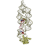
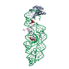
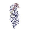
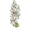
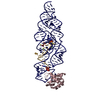
| Title | Crystal structure of the Bacillus anthracis glmS ribozyme bound to Glc6P | |||||||||
 Components Components |
| |||||||||
 Keywords Keywords | RNA binding protein/RNA /  catalytic RNA / RNA binding protein-RNA complex catalytic RNA / RNA binding protein-RNA complex | |||||||||
| Function / homology |  Function and homology information Function and homology informationU1 snRNP binding / U1 snRNP / U1 snRNA binding / U4/U6 x U5 tri-snRNP complex / mRNA Splicing - Major Pathway / spliceosomal complex / mRNA splicing, via spliceosome / DNA binding / RNA binding / nucleoplasm ...U1 snRNP binding / U1 snRNP / U1 snRNA binding / U4/U6 x U5 tri-snRNP complex / mRNA Splicing - Major Pathway / spliceosomal complex / mRNA splicing, via spliceosome / DNA binding / RNA binding / nucleoplasm / identical protein binding / nucleusSimilarity search - Function | |||||||||
| Biological species |  HOMO SAPIENS (human) HOMO SAPIENS (human) | |||||||||
| Method | X-RAY DIFFRACTION / SYNCHROTRON / MOLECULAR REPLACEMENT / Resolution: 2.85 Å | |||||||||
 Authors Authors | Strobel, S.A. / Cochrane, J.C. / Lipchock, S.V. / Smith, K.D. | |||||||||
 Citation Citation | Journal: Biochemistry / Year: 2009 Title: Structural and chemical basis for glucosamine 6-phosphate binding and activation of the glmS ribozyme Authors: Cochrane, J.C. / Lipchock, S.V. / Smith, K.D. / Strobel, S.A. | |||||||||
| History |
|
- Structure visualization
| Structure viewer | Molecule: MolmilJmol/JSmol |
|---|
- Downloads & links
-Download
| PDBx/mmCIF format | 3l3c.cif.gz | 409 KB | Display | PDBx/mmCIF format |
|---|---|---|---|---|
| PDB format | pdb3l3c.ent.gz | 317.3 KB | Display | PDB format |
| PDBx/mmJSON format | 3l3c.json.gz | Tree view | PDBx/mmJSON format | |
| Others |  Other downloads Other downloads |
-Validation report
| Arichive directory | https://data.pdbj.org/pub/pdb/validation_reports/l3/3l3cftp://data.pdbj.org/pub/pdb/validation_reports/l3/3l3c | HTTPS FTP |
|---|
-Related structure data


-Links
 PDBj
PDBj


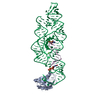
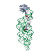
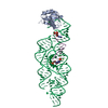
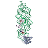
- Assembly
| Deposited unit | 
| ||||||||
|---|---|---|---|---|---|---|---|---|---|
| 1 | 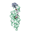
| ||||||||
| 2 | 
| ||||||||
| 3 | 
| ||||||||
| 4 | 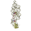
| ||||||||
| Unit cell |
| ||||||||
| Details | The biological unit is a ternary complex and there are four biological units in the asymmetric unit (chains a,e&p, chains b,f&q, chains c,g&r, and chains d,h&s |
-Components
-RNA chain , 2 types, 8 molecules EFGHPQRS
| #2: RNA chain | Mass: 4177.608 Da / Num. of mol.: 4 / Mutation: 2'-OH at A-1 / Source method: obtained synthetically / Details: synthesized at Dharmacon #3: RNA chain | Mass: 45624.859 Da / Num. of mol.: 4 / Source method: obtained synthetically / Details: in vitro transcribed from a DNA template |
|---|
-Protein / Sugars , 2 types, 8 molecules ABCD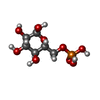
| #1: Protein | Mass: 10451.204 Da / Num. of mol.: 4 / Fragment: RNA BINDING DOMAIN / Mutation: Y31H,Q36R Source method: isolated from a genetically manipulated source Source: (gene. exp.) HOMO SAPIENS (human) / Gene: SNRPA / Plasmid: pET11 / Production host:  Escherichia coli (E. coli) / Strain (production host): BL21 / References: UniProt: P09012 Escherichia coli (E. coli) / Strain (production host): BL21 / References: UniProt: P09012#5: Sugar | ChemComp-G6P /  Type: D-saccharide, alpha linking / Mass: 260.136 Da / Num. of mol.: 4 Type: D-saccharide, alpha linking / Mass: 260.136 Da / Num. of mol.: 4Source method: isolated from a genetically manipulated source Formula: C6H13O9P |
|---|
-Non-polymers , 2 types, 54 molecules 

| #4: Chemical | ChemComp-MG /  Mass: 24.305 Da / Num. of mol.: 16 / Source method: obtained synthetically / Formula: Mg Mass: 24.305 Da / Num. of mol.: 16 / Source method: obtained synthetically / Formula: Mg#6: Water | ChemComp-HOH / | WaterMass: 18.015 Da / Num. of mol.: 38 / Source method: isolated from a natural source / Formula: H2O |
|---|
-Experimental details
-Experiment
| Experiment | Method: X-RAY DIFFRACTION / Number of used crystals: 1 |
|---|
- Sample preparation
| Crystal | Density Matthews: 2.44 Å3/Da / Density % sol: 49.58 % |
|---|---|
| Crystal grow | Temperature: 298 K / Method: vapor diffusion, sitting drop / pH: 6.8 Details: 11% PEG 8000, 9% DMSO, 0.02M SODIUM CACODYLATE, 0.02M MAGNESIUM CHLORIDE, 0.15M POTASSIUM CHLORIDE, pH 6.8, vapor diffusion, sitting drops, temperature 298K |
-Data collection
| Diffraction | Mean temperature: 100 K |
|---|---|
| Diffraction source | Source: SYNCHROTRON / Site: NSLS  / Beamline: X25 / Wavelength: 1.1 Å / Beamline: X25 / Wavelength: 1.1 Å |
| Detector | Type: ADSC QUANTUM 315 / Detector: CCD / Date: Oct 5, 2007 |
| Radiation | Protocol: SINGLE WAVELENGTH / Monochromatic (M) / Laue (L): M / Scattering type: x-ray |
| Radiation wavelength | Wavelength: 1.1 Å / Relative weight: 1 |
| Reflection | Resolution: 2.85→50 Å / Num. all: 52648 / Num. obs: 52648 / % possible obs: 98.1 % / Observed criterion σ(F): 0 / Observed criterion σ(I): 0 / Redundancy: 4 % / Rmerge(I) obs: 0.062 / Χ2: 1.029 / Net I/σ(I): 14.4 |
| Reflection shell | Resolution: 2.85→2.95 Å / Redundancy: 3.2 % / Rmerge(I) obs: 0.832 / Num. unique all: 4552 / Χ2: 1.026 / % possible all: 85.5 |
- Processing
| Software |
| |||||||||||||||||||||||||||||||||||||||||||||||||||||||||||||||||
|---|---|---|---|---|---|---|---|---|---|---|---|---|---|---|---|---|---|---|---|---|---|---|---|---|---|---|---|---|---|---|---|---|---|---|---|---|---|---|---|---|---|---|---|---|---|---|---|---|---|---|---|---|---|---|---|---|---|---|---|---|---|---|---|---|---|---|
| Refinement | Method to determine structure: MOLECULAR REPLACEMENT / Resolution: 2.85→46.93 Å / Cor.coef. Fo:Fc: 0.93 / Cor.coef. Fo:Fc free: 0.904 / Occupancy max: 1 / Occupancy min: 1 / SU B: 23.037 / SU ML: 0.447 / Cross valid method: THROUGHOUT / σ(F): 0 / ESU R Free: 0.502 / Stereochemistry target values: MAXIMUM LIKELIHOOD Details: HYDROGENS HAVE BEEN ADDED IN THE RIDING POSITIONS U VALUES : REFINED INDIVIDUALLY
| |||||||||||||||||||||||||||||||||||||||||||||||||||||||||||||||||
| Solvent computation | Ion probe radii: 0.8 Å / Shrinkage radii: 0.8 Å / VDW probe radii: 1.4 Å / Solvent model: MASK | |||||||||||||||||||||||||||||||||||||||||||||||||||||||||||||||||
| Displacement parameters | Biso max: 162.32 Å2 / Biso mean: 83.982 Å2 / Biso min: 33.08 Å2
| |||||||||||||||||||||||||||||||||||||||||||||||||||||||||||||||||
| Refinement step | Cycle: LAST / Resolution: 2.85→46.93 Å
| |||||||||||||||||||||||||||||||||||||||||||||||||||||||||||||||||
| Refine LS restraints |
| |||||||||||||||||||||||||||||||||||||||||||||||||||||||||||||||||
| LS refinement shell | Resolution: 2.847→2.921 Å / Total num. of bins used: 20
|
