 Movie
Movie Controller


+ Open data

- Basic information
| Entry | Database: PDB / ID: 5glz | ||||||
|---|---|---|---|---|---|---|---|
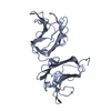
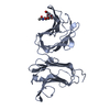
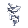
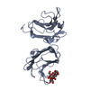
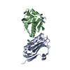
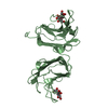
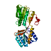
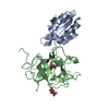
| Title | Tl-gal with Glucose | ||||||
 Components Components | galectin | ||||||
 Keywords Keywords | SUGAR BINDING PROTEIN / Carbohydrates / Tl-galectin / Anti-inflammation | ||||||
| Function / homology |  Function and homology information Function and homology information | ||||||
| Biological species |  Toxascaris leonina (invertebrata) Toxascaris leonina (invertebrata) | ||||||
| Method | X-RAY DIFFRACTION / SYNCHROTRON / MOLECULAR REPLACEMENT / Resolution: 2 Å | ||||||
 Authors Authors | Jang, S.B. / Hwang, E.Y. | ||||||
| Funding support |  Korea, Republic Of, 1items Korea, Republic Of, 1items
| ||||||
 Citation Citation | Journal: J. Biol. Chem. / Year: 2016 Title: Structural Basis for Carbohydrate Recognition and Anti-inflammatory Modulation by Gastrointestinal Nematode Parasite Toxascaris leonina Galectin Authors: Hwang, E.Y. / Jeong, M.S. / Park, S.K. / Ha, S.C. / Yu, H.S. / Jang, S.B. #1: Journal: Acta Crystallogr.,Sect.D / Year: 2013Title: Structure of full-length Toxascaris leonina galectin with two carbohydrate-recognition domains Authors: Jeong, M.S. / Hwang, H.G. / Yu, H.S. / Jang, S.B. | ||||||
| History |
|
- Structure visualization
| Structure viewer | Molecule: MolmilJmol/JSmol |
|---|
- Downloads & links
-Download
| PDBx/mmCIF format | 5glz.cif.gz | 243.4 KB | Display | PDBx/mmCIF format |
|---|---|---|---|---|
| PDB format | pdb5glz.ent.gz | 194.7 KB | Display | PDB format |
| PDBx/mmJSON format | 5glz.json.gz | Tree view | PDBx/mmJSON format | |
| Others |  Other downloads Other downloads |
-Validation report
| Arichive directory | https://data.pdbj.org/pub/pdb/validation_reports/gl/5glzftp://data.pdbj.org/pub/pdb/validation_reports/gl/5glz | HTTPS FTP |
|---|
-Related structure data
| Related structure data | 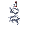 5gltC  5gluC  5glvC  5glwC 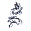 5gm0C  4hl0S C: citing same article ( S: Starting model for refinement |
|---|---|
| Similar structure data |


-Links
 PDBj
PDBj


- Assembly
| Deposited unit | 
| ||||||||
|---|---|---|---|---|---|---|---|---|---|
| 1 | 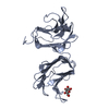
| ||||||||
| 2 | 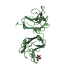
| ||||||||
| 3 | 
| ||||||||
| 4 | 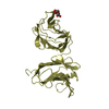
| ||||||||
| Unit cell |
|
-Components
| #1: Protein | Mass: 32324.168 Da / Num. of mol.: 4 Source method: isolated from a genetically manipulated source Source: (gene. exp.) Toxascaris leonina (invertebrata) / Production host:  Escherichia coli (E. coli) / References: UniProt: A0A1L1QJZ7*PLUS Escherichia coli (E. coli) / References: UniProt: A0A1L1QJZ7*PLUS#2: Sugar | ChemComp-BGC / Glucose  Type: D-saccharide, beta linking / Mass: 180.156 Da / Num. of mol.: 4 / Source method: obtained synthetically / Formula: C6H12O6 Type: D-saccharide, beta linking / Mass: 180.156 Da / Num. of mol.: 4 / Source method: obtained synthetically / Formula: C6H12O6#3: Water | ChemComp-HOH / | Water Mass: 18.015 Da / Num. of mol.: 697 / Source method: isolated from a natural source / Formula: H2O Mass: 18.015 Da / Num. of mol.: 697 / Source method: isolated from a natural source / Formula: H2O |
|---|
-Experimental details
-Experiment
| Experiment | Method: X-RAY DIFFRACTION / Number of used crystals: 1 |
|---|
- Sample preparation
| Crystal | Density Matthews: 3.46 Å3/Da / Density % sol: 64.44 % |
|---|---|
| Crystal grow | Temperature: 293 K / Method: vapor diffusion, hanging drop Details: 10% polyethylene glycol 8000, 100mM Na/K phosphate (pH 6.2), 200mM NaCl |
-Data collection
| Diffraction | Mean temperature: 100 K |
|---|---|
| Diffraction source | Source: SYNCHROTRON / Site: PAL/PLS / Beamline: 7A (6B, 6C1) / Wavelength: 0.987 Å |
| Detector | Type: MAC Science DIP-320 / Detector: IMAGE PLATE / Date: Jun 24, 2014 |
| Radiation | Protocol: SINGLE WAVELENGTH / Monochromatic (M) / Laue (L): M / Scattering type: x-ray |
| Radiation wavelength | Wavelength: 0.987 Å / Relative weight: 1 |
| Reflection | Resolution: 2→30 Å / Num. obs: 114308 / % possible obs: 96.4 % / Redundancy: 1.9 % / Rmerge(I) obs: 0.055 / Net I/σ(I): 12.6 |
| Reflection shell | Resolution: 2→2.07 Å / Rmerge(I) obs: 0.224 |
- Processing
| Software |
| ||||||||||||||||||||
|---|---|---|---|---|---|---|---|---|---|---|---|---|---|---|---|---|---|---|---|---|---|
| Refinement | Method to determine structure: MOLECULAR REPLACEMENT Starting model: 4HL0 Resolution: 2→30 Å / Cross valid method: FREE R-VALUE / σ(F): 0
| ||||||||||||||||||||
| Solvent computation | Bsol: 59.8004 Å2 | ||||||||||||||||||||
| Displacement parameters | Biso max: 95.45 Å2 / Biso mean: 27.1991 Å2 / Biso min: 6.15 Å2
| ||||||||||||||||||||
| Refinement step | Cycle: LAST / Resolution: 2→30 Å
| ||||||||||||||||||||
| Refine LS restraints |
| ||||||||||||||||||||
| Xplor file |
|
