 Movie
Movie Controller

[English] 日本語
 Yorodumi
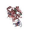
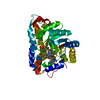
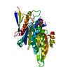
- PDB-5gjf: Crystal structure of human TAK1/TAB1 fusion protein in complex wi... -
+ Open data

- Basic information
| Entry | Database: PDB / ID: 5gjf | ||||||
|---|---|---|---|---|---|---|---|
| Title | Crystal structure of human TAK1/TAB1 fusion protein in complex with ligand 3 | ||||||
 Components Components | TAK1 kinase - TAB1 chimera fusion protein | ||||||
 Keywords Keywords | TRANSFERASE/TRANSFERASE INHIBITOR / Transferase-Transferase inhibitor complex / TAK1-TAB1 kinase | ||||||
| Function / homology |  Function and homology information Function and homology informationnucleotide-binding domain, leucine rich repeat containing receptor signaling pathway / positive regulation of cGAS/STING signaling pathway / histone kinase activity /  MAP kinase kinase kinase kinase activity / linear polyubiquitin binding / interleukin-17A-mediated signaling pathway / cardiac septum development / I-kappaB phosphorylation / mitogen-activated protein kinase kinase kinase / interleukin-33-mediated signaling pathway ...nucleotide-binding domain, leucine rich repeat containing receptor signaling pathway / positive regulation of cGAS/STING signaling pathway / histone kinase activity / MAP kinase kinase kinase kinase activity / linear polyubiquitin binding / interleukin-17A-mediated signaling pathway / cardiac septum development / I-kappaB phosphorylation / mitogen-activated protein kinase kinase kinase / interleukin-33-mediated signaling pathway / toll-like receptor 3 signaling pathway / type II transforming growth factor beta receptor binding / TRIF-dependent toll-like receptor signaling pathway / activation of NF-kappaB-inducing kinase activity / coronary vasculature development / ATAC complex / positive regulation of vascular associated smooth muscle cell migration / cellular response to angiotensin / anoikis / aorta development / MyD88-dependent toll-like receptor signaling pathway / interleukin-1-mediated signaling pathway / toll-like receptor 4 signaling pathway / protein serine/threonine phosphatase activity / protein serine/threonine kinase binding / mitogen-activated protein kinase p38 binding / cytoplasmic pattern recognition receptor signaling pathway / p38MAPK cascade / non-canonical NF-kappaB signal transduction / Fc-epsilon receptor signaling pathway / stimulatory C-type lectin receptor signaling pathway / positive regulation of macroautophagy / positive regulation of cell size / enzyme activator activity / MAP kinase kinase kinase activity / MAP kinase activity / canonical NF-kappaB signal transduction / stress-activated MAPK cascade / positive regulation of cell cycle / heart morphogenesis / positive regulation of JUN kinase activity / JNK cascade / IRAK2 mediated activation of TAK1 complex / Alpha-protein kinase 1 signaling pathway / positive regulation of vascular associated smooth muscle cell proliferation / IRAK2 mediated activation of TAK1 complex upon TLR7/8 or 9 stimulation / TICAM1,TRAF6-dependent induction of TAK1 complex / TRAF6-mediated induction of TAK1 complex within TLR4 complex / positive regulation of interleukin-2 production / protein serine/threonine kinase activator activity / transforming growth factor beta receptor signaling pathway / JNK (c-Jun kinases) phosphorylation and activation mediated by activated human TAK1 / TNFR1-induced NF-kappa-B signaling pathway / activated TAK1 mediates p38 MAPK activation / Activation of NF-kappaB in B cells / lung development / TAK1-dependent IKK and NF-kappa-B activation / NOD1/2 Signaling Pathway / positive regulation of protein serine/threonine kinase activity / transcription coactivator binding / receptor tyrosine kinase binding / CLEC7A (Dectin-1) signaling / positive regulation of T cell cytokine production / FCERI mediated NF-kB activation / Interleukin-1 signaling / positive regulation of non-canonical NF-kappaB signal transduction / MAPK cascade / Downstream TCR signaling / Ca2+ pathway / cellular response to tumor necrosis factor / cellular response to hypoxia / T cell receptor signaling pathway / scaffold protein binding / positive regulation of canonical NF-kappaB signal transduction / DNA-binding transcription factor binding / in utero embryonic development / positive regulation of MAPK cascade / molecular adaptor activity / endosome membrane / Ub-specific processing proteases / nuclear speck / defense response to bacterium / immune response / inflammatory response / negative regulation of gene expression / protein serine kinase activity / protein serine/threonine kinase activity / ubiquitin protein ligase binding / protein-containing complex binding / endoplasmic reticulum membrane / SARS-CoV-2 activates/modulates innate and adaptive immune responses / magnesium ion binding / endoplasmic reticulum / protein-containing complex / ATP binding / identical protein binding / nucleus / plasma membrane / cytosol / cytoplasm MAP kinase kinase kinase kinase activity / linear polyubiquitin binding / interleukin-17A-mediated signaling pathway / cardiac septum development / I-kappaB phosphorylation / mitogen-activated protein kinase kinase kinase / interleukin-33-mediated signaling pathway ...nucleotide-binding domain, leucine rich repeat containing receptor signaling pathway / positive regulation of cGAS/STING signaling pathway / histone kinase activity / MAP kinase kinase kinase kinase activity / linear polyubiquitin binding / interleukin-17A-mediated signaling pathway / cardiac septum development / I-kappaB phosphorylation / mitogen-activated protein kinase kinase kinase / interleukin-33-mediated signaling pathway / toll-like receptor 3 signaling pathway / type II transforming growth factor beta receptor binding / TRIF-dependent toll-like receptor signaling pathway / activation of NF-kappaB-inducing kinase activity / coronary vasculature development / ATAC complex / positive regulation of vascular associated smooth muscle cell migration / cellular response to angiotensin / anoikis / aorta development / MyD88-dependent toll-like receptor signaling pathway / interleukin-1-mediated signaling pathway / toll-like receptor 4 signaling pathway / protein serine/threonine phosphatase activity / protein serine/threonine kinase binding / mitogen-activated protein kinase p38 binding / cytoplasmic pattern recognition receptor signaling pathway / p38MAPK cascade / non-canonical NF-kappaB signal transduction / Fc-epsilon receptor signaling pathway / stimulatory C-type lectin receptor signaling pathway / positive regulation of macroautophagy / positive regulation of cell size / enzyme activator activity / MAP kinase kinase kinase activity / MAP kinase activity / canonical NF-kappaB signal transduction / stress-activated MAPK cascade / positive regulation of cell cycle / heart morphogenesis / positive regulation of JUN kinase activity / JNK cascade / IRAK2 mediated activation of TAK1 complex / Alpha-protein kinase 1 signaling pathway / positive regulation of vascular associated smooth muscle cell proliferation / IRAK2 mediated activation of TAK1 complex upon TLR7/8 or 9 stimulation / TICAM1,TRAF6-dependent induction of TAK1 complex / TRAF6-mediated induction of TAK1 complex within TLR4 complex / positive regulation of interleukin-2 production / protein serine/threonine kinase activator activity / transforming growth factor beta receptor signaling pathway / JNK (c-Jun kinases) phosphorylation and activation mediated by activated human TAK1 / TNFR1-induced NF-kappa-B signaling pathway / activated TAK1 mediates p38 MAPK activation / Activation of NF-kappaB in B cells / lung development / TAK1-dependent IKK and NF-kappa-B activation / NOD1/2 Signaling Pathway / positive regulation of protein serine/threonine kinase activity / transcription coactivator binding / receptor tyrosine kinase binding / CLEC7A (Dectin-1) signaling / positive regulation of T cell cytokine production / FCERI mediated NF-kB activation / Interleukin-1 signaling / positive regulation of non-canonical NF-kappaB signal transduction / MAPK cascade / Downstream TCR signaling / Ca2+ pathway / cellular response to tumor necrosis factor / cellular response to hypoxia / T cell receptor signaling pathway / scaffold protein binding / positive regulation of canonical NF-kappaB signal transduction / DNA-binding transcription factor binding / in utero embryonic development / positive regulation of MAPK cascade / molecular adaptor activity / endosome membrane / Ub-specific processing proteases / nuclear speck / defense response to bacterium / immune response / inflammatory response / negative regulation of gene expression / protein serine kinase activity / protein serine/threonine kinase activity / ubiquitin protein ligase binding / protein-containing complex binding / endoplasmic reticulum membrane / SARS-CoV-2 activates/modulates innate and adaptive immune responses / magnesium ion binding / endoplasmic reticulum / protein-containing complex / ATP binding / identical protein binding / nucleus / plasma membrane / cytosol / cytoplasmSimilarity search - Function | ||||||
| Biological species |  Homo sapiens (human) Homo sapiens (human) | ||||||
| Method | X-RAY DIFFRACTION / SYNCHROTRON / MOLECULAR REPLACEMENT / molecular replacement / Resolution: 2.89 Å | ||||||
 Authors Authors | Irie, M. / Nakamura, M. / Fukami, T.A. / Matsuura, T. / Morishima, K. | ||||||
 Citation Citation | Journal: Chem.Pharm.Bull. / Year: 2016 Title: Development of a Method for Converting a TAK1 Type I Inhibitor into a Type II or c-Helix-Out Inhibitor by Structure-Based Drug Design (SBDD) Authors: Muraoka, T. / Ide, M. / Irie, M. / Morikami, K. / Miura, T. / Nishihara, M. / Kashiwagi, H. | ||||||
| History |
|
- Structure visualization
| Structure viewer | Molecule: MolmilJmol/JSmol |
|---|
- Downloads & links
-Download
| PDBx/mmCIF format | 5gjf.cif.gz | 76.8 KB | Display | PDBx/mmCIF format |
|---|---|---|---|---|
| PDB format | pdb5gjf.ent.gz | 54 KB | Display | PDB format |
| PDBx/mmJSON format | 5gjf.json.gz | Tree view | PDBx/mmJSON format | |
| Others |  Other downloads Other downloads |
-Validation report
| Arichive directory | https://data.pdbj.org/pub/pdb/validation_reports/gj/5gjfftp://data.pdbj.org/pub/pdb/validation_reports/gj/5gjf | HTTPS FTP |
|---|
-Related structure data
| Related structure data |  5gjdC  5gjgC  2evaS C: citing same article ( S: Starting model for refinement |
|---|---|
| Similar structure data |


-Links
 PDBj
PDBj


- Assembly
| Deposited unit | 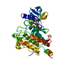
| ||||||||
|---|---|---|---|---|---|---|---|---|---|
| 1 |
| ||||||||
| Unit cell |
|
-Components
| #1: Protein | Mass: 35530.953 Da / Num. of mol.: 1 / Fragment: UNP residues 31-303,UNP residues 468-504 Source method: isolated from a genetically manipulated source Details: fusion of Mitogen-activated protein kinase kinase kinase 7,TGF-beta-activated kinase 1 and MAP3K7-binding protein 1 Source: (gene. exp.) Homo sapiens (human) / Gene: MAP3K7, TAK1, TAB1, MAP3K7IP1 / Cell line (production host): Sf9 / Production host:   Spodoptera frugiperda (fall armyworm) Spodoptera frugiperda (fall armyworm)References: UniProt: O43318, UniProt: Q15750, mitogen-activated protein kinase kinase kinase |
|---|---|
| #2: Chemical | ChemComp-6V4 /   Mass: 598.715 Da / Num. of mol.: 1 / Source method: obtained synthetically / Formula: C32H34N6O4S Mass: 598.715 Da / Num. of mol.: 1 / Source method: obtained synthetically / Formula: C32H34N6O4S |
| #3: Water | ChemComp-HOH / Water Mass: 18.015 Da / Num. of mol.: 13 / Source method: isolated from a natural source / Formula: H2O Mass: 18.015 Da / Num. of mol.: 13 / Source method: isolated from a natural source / Formula: H2O |
-Experimental details
-Experiment
| Experiment | Method: X-RAY DIFFRACTION / Number of used crystals: 1 |
|---|
- Sample preparation
| Crystal | Density Matthews: 3.84 Å3/Da / Density % sol: 67.99 % |
|---|---|
| Crystal grow | Temperature: 294 K / Method: vapor diffusion, hanging drop / pH: 7.5 Details: 1.94M sodium potassium phosphate, 20%(v/v) Glycerol as cryoprotectant |
-Data collection
| Diffraction | Mean temperature: 95 K | ||||||||||||||||||||||||||||||||||||||||||||||||||||||||||||||||||||||||||||||||||||||||||||||||||||||||||||||||||||||||||||||
|---|---|---|---|---|---|---|---|---|---|---|---|---|---|---|---|---|---|---|---|---|---|---|---|---|---|---|---|---|---|---|---|---|---|---|---|---|---|---|---|---|---|---|---|---|---|---|---|---|---|---|---|---|---|---|---|---|---|---|---|---|---|---|---|---|---|---|---|---|---|---|---|---|---|---|---|---|---|---|---|---|---|---|---|---|---|---|---|---|---|---|---|---|---|---|---|---|---|---|---|---|---|---|---|---|---|---|---|---|---|---|---|---|---|---|---|---|---|---|---|---|---|---|---|---|---|---|---|
| Diffraction source | Source: SYNCHROTRON / Site: Photon Factory  / Beamline: BL-17A / Wavelength: 1 Å / Beamline: BL-17A / Wavelength: 1 Å | ||||||||||||||||||||||||||||||||||||||||||||||||||||||||||||||||||||||||||||||||||||||||||||||||||||||||||||||||||||||||||||||
| Detector | Type: ADSC QUANTUM 270 / Detector: CCD / Date: Dec 14, 2009 | ||||||||||||||||||||||||||||||||||||||||||||||||||||||||||||||||||||||||||||||||||||||||||||||||||||||||||||||||||||||||||||||
| Radiation | Protocol: SINGLE WAVELENGTH / Monochromatic (M) / Laue (L): M / Scattering type: x-ray | ||||||||||||||||||||||||||||||||||||||||||||||||||||||||||||||||||||||||||||||||||||||||||||||||||||||||||||||||||||||||||||||
| Radiation wavelength | Wavelength: 1 Å / Relative weight: 1 | ||||||||||||||||||||||||||||||||||||||||||||||||||||||||||||||||||||||||||||||||||||||||||||||||||||||||||||||||||||||||||||||
| Reflection | Resolution: 2.89→48.441 Å / Num. all: 12633 / Num. obs: 12633 / % possible obs: 99.8 % / Redundancy: 7.1 % / Rpim(I) all: 0.057 / Rrim(I) all: 0.153 / Rsym value: 0.142 / Net I/av σ(I): 5.182 / Net I/σ(I): 12.7 / Num. measured all: 90311 | ||||||||||||||||||||||||||||||||||||||||||||||||||||||||||||||||||||||||||||||||||||||||||||||||||||||||||||||||||||||||||||||
| Reflection shell |
|
-Phasing
| Phasing | Method: molecular replacement |
|---|
- Processing
| Software |
| |||||||||||||||||||||||||||||||||||||||||||||||||||||||||||||||||||||||||||
|---|---|---|---|---|---|---|---|---|---|---|---|---|---|---|---|---|---|---|---|---|---|---|---|---|---|---|---|---|---|---|---|---|---|---|---|---|---|---|---|---|---|---|---|---|---|---|---|---|---|---|---|---|---|---|---|---|---|---|---|---|---|---|---|---|---|---|---|---|---|---|---|---|---|---|---|---|
| Refinement | Method to determine structure: MOLECULAR REPLACEMENT Starting model: 2EVA Resolution: 2.89→48.44 Å / Cor.coef. Fo:Fc: 0.936 / Cor.coef. Fo:Fc free: 0.916 / SU B: 12.559 / SU ML: 0.233 / Cross valid method: THROUGHOUT / σ(F): 0 / ESU R: 0.56 / ESU R Free: 0.305 Details: HYDROGENS HAVE BEEN ADDED IN THE RIDING POSITIONS U VALUES : REFINED INDIVIDUALLY
| |||||||||||||||||||||||||||||||||||||||||||||||||||||||||||||||||||||||||||
| Solvent computation | Ion probe radii: 0.8 Å / Shrinkage radii: 0.8 Å / VDW probe radii: 1.2 Å | |||||||||||||||||||||||||||||||||||||||||||||||||||||||||||||||||||||||||||
| Displacement parameters | Biso max: 140.77 Å2 / Biso mean: 64.998 Å2 / Biso min: 23.94 Å2
| |||||||||||||||||||||||||||||||||||||||||||||||||||||||||||||||||||||||||||
| Refinement step | Cycle: final / Resolution: 2.89→48.44 Å
| |||||||||||||||||||||||||||||||||||||||||||||||||||||||||||||||||||||||||||
| Refine LS restraints |
| |||||||||||||||||||||||||||||||||||||||||||||||||||||||||||||||||||||||||||
| LS refinement shell | Resolution: 2.89→2.965 Å / Total num. of bins used: 20
|
