 Movie
Movie Controller


+ Open data

- Basic information
| Entry | Database: PDB / ID: 5gij | |||||||||
|---|---|---|---|---|---|---|---|---|---|---|
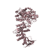
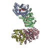
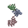
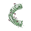
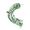
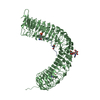
| Title | Crystal structure of TDR-TDIF complex | |||||||||
 Components Components |
| |||||||||
 Keywords Keywords |  SIGNALING PROTEIN / LRR / TDR / TDIF / PXY / LRR-RK SIGNALING PROTEIN / LRR / TDR / TDIF / PXY / LRR-RK | |||||||||
| Function / homology |  Function and homology information Function and homology informationprocambium histogenesis / phloem development / xylem development / axillary shoot meristem initiation / maintenance of root meristem identity / cell-cell signaling involved in cell fate commitment / phloem or xylem histogenesis / secondary shoot formation / apoplast / receptor serine/threonine kinase binding ...procambium histogenesis / phloem development / xylem development / axillary shoot meristem initiation / maintenance of root meristem identity / cell-cell signaling involved in cell fate commitment / phloem or xylem histogenesis / secondary shoot formation / apoplast / receptor serine/threonine kinase binding / regulation of cell differentiation / non-specific serine/threonine protein kinase / cell division / phosphorylation / protein serine kinase activity / protein serine/threonine kinase activity / ATP binding / plasma membraneSimilarity search - Function | |||||||||
| Biological species |  Arabidopsis thaliana (thale cress) Arabidopsis thaliana (thale cress) | |||||||||
| Method | X-RAY DIFFRACTION / SYNCHROTRON / MOLECULAR REPLACEMENT / Resolution: 3 Å | |||||||||
 Authors Authors | Morita, J. / Kato, K. / Ishitani, R. / Nishimasu, H. / Nureki, O. | |||||||||
 Citation Citation | Journal: Nat Commun / Year: 2016 Title: Crystal structure of the plant receptor-like kinase TDR in complex with the TDIF peptide Authors: Morita, J. / Kato, K. / Nakane, T. / Kondo, Y. / Fukuda, H. / Nishimasu, H. / Ishitani, R. / Nureki, O. | |||||||||
| History |
|
- Structure visualization
| Structure viewer | Molecule: MolmilJmol/JSmol |
|---|
- Downloads & links
-Download
| PDBx/mmCIF format | 5gij.cif.gz | 141.6 KB | Display | PDBx/mmCIF format |
|---|---|---|---|---|
| PDB format | pdb5gij.ent.gz | 109.4 KB | Display | PDB format |
| PDBx/mmJSON format | 5gij.json.gz | Tree view | PDBx/mmJSON format | |
| Others |  Other downloads Other downloads |
-Validation report
| Arichive directory | https://data.pdbj.org/pub/pdb/validation_reports/gi/5gijftp://data.pdbj.org/pub/pdb/validation_reports/gi/5gij | HTTPS FTP |
|---|
-Related structure data
| Related structure data | 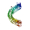 4mnaS S: Starting model for refinement |
|---|---|
| Similar structure data |


-Links
 PDBj
PDBj


- Assembly
| Deposited unit | 
| ||||||||
|---|---|---|---|---|---|---|---|---|---|
| 1 |
| ||||||||
| Unit cell |
|
-Components
-Protein / Protein/peptide , 2 types, 2 molecules BD
| #1: Protein | Mass: 66735.195 Da / Num. of mol.: 1 / Fragment: extracellular domain, UNP residues 31-631 / Mutation: C259A, C540S Source method: isolated from a genetically manipulated source Source: (gene. exp.) Arabidopsis thaliana (thale cress) / Gene: TDR, PXY, At5g61480, MCI2.4 / Plasmid: pFastBac1 / Cell line (production host): Sf9 / Production host:   Spodoptera frugiperda (fall armyworm) Spodoptera frugiperda (fall armyworm)References: UniProt: Q9FII5, non-specific serine/threonine protein kinase |
|---|---|
| #2: Protein/peptide | / Tracheary element differentiation inhibitory factor-like protein / TDIF-like protein Mass: 1280.322 Da / Num. of mol.: 1 / Source method: obtained synthetically / Source: (synth.) Arabidopsis thaliana (thale cress) / References: UniProt: Q84W98 |
-Sugars , 5 types, 11 molecules 
| #3: Polysaccharide | alpha-D-mannopyranose-(1-4)-2-acetamido-2-deoxy-beta-D-glucopyranose-(1-4)-2-acetamido-2-deoxy-beta- ...alpha-D-mannopyranose-(1-4)-2-acetamido-2-deoxy-beta-D-glucopyranose-(1-4)-2-acetamido-2-deoxy-beta-D-glucopyranose / Mass: 586.542 Da / Num. of mol.: 1 Source method: isolated from a genetically manipulated source | ||||||
|---|---|---|---|---|---|---|---|
| #4: Polysaccharide | 2-acetamido-2-deoxy-beta-D-glucopyranose-(1-4)-2-acetamido-2-deoxy-beta-D-glucopyranose / Mass: 424.401 Da / Num. of mol.: 4Source method: isolated from a genetically manipulated source #5: Polysaccharide | alpha-D-mannopyranose-(1-6)-alpha-D-mannopyranose-(1-4)-2-acetamido-2-deoxy-beta-D-glucopyranose-(1- ...alpha-D-mannopyranose-(1-6)-alpha-D-mannopyranose-(1-4)-2-acetamido-2-deoxy-beta-D-glucopyranose-(1-4)-2-acetamido-2-deoxy-beta-D-glucopyranose | / Mass: 748.682 Da / Num. of mol.: 1Source method: isolated from a genetically manipulated source #6: Polysaccharide | alpha-D-mannopyranose-(1-6)-alpha-D-mannopyranose-(1-4)-2-acetamido-2-deoxy-beta-D-glucopyranose-(1- ...alpha-D-mannopyranose-(1-6)-alpha-D-mannopyranose-(1-4)-2-acetamido-2-deoxy-beta-D-glucopyranose-(1-4)-[alpha-L-fucopyranose-(1-6)]2-acetamido-2-deoxy-beta-D-glucopyranose | / Mass: 894.823 Da / Num. of mol.: 1Source method: isolated from a genetically manipulated source #7: Sugar | ChemComp-NAG / N-Acetylglucosamine Type: D-saccharide, beta linking / Mass: 221.208 Da / Num. of mol.: 4 Type: D-saccharide, beta linking / Mass: 221.208 Da / Num. of mol.: 4Source method: isolated from a genetically manipulated source Formula: C8H15NO6 |
-Experimental details
-Experiment
| Experiment | Method: X-RAY DIFFRACTION / Number of used crystals: 1 |
|---|
- Sample preparation
| Crystal | Density Matthews: 8.62 Å3/Da / Density % sol: 85.73 % |
|---|---|
| Crystal grow | Temperature: 293 K / Method: vapor diffusion Details: 4M sodium nitrate, 0.1M sodium acetate trihydrate, pH 4.8, 200mM ammonium sulfate |
-Data collection
| Diffraction | Mean temperature: 100 K |
|---|---|
| Diffraction source | Source: SYNCHROTRON / Site: SLS  / Beamline: X06SA / Wavelength: 1 Å / Beamline: X06SA / Wavelength: 1 Å |
| Detector | Type: DECTRIS PILATUS 6M-F / Detector: PIXEL / Date: Mar 13, 2015 |
| Radiation | Protocol: SINGLE WAVELENGTH / Monochromatic (M) / Laue (L): M / Scattering type: x-ray |
| Radiation wavelength | Wavelength: 1 Å / Relative weight: 1 |
| Reflection | Resolution: 3→102.93 Å / Num. obs: 47698 / % possible obs: 99.8 % / Redundancy: 9.7 % / Rmerge(I) obs: 0.152 / Net I/σ(I): 8.8 |
| Reflection shell | Resolution: 3→3.11 Å |
- Processing
| Software |
| ||||||||||||||||||||||||||||||||||||||||||||||||||||||||||||||||||||||||||||||||||||||||||||||||||||||||||||||||||||||||||||||
|---|---|---|---|---|---|---|---|---|---|---|---|---|---|---|---|---|---|---|---|---|---|---|---|---|---|---|---|---|---|---|---|---|---|---|---|---|---|---|---|---|---|---|---|---|---|---|---|---|---|---|---|---|---|---|---|---|---|---|---|---|---|---|---|---|---|---|---|---|---|---|---|---|---|---|---|---|---|---|---|---|---|---|---|---|---|---|---|---|---|---|---|---|---|---|---|---|---|---|---|---|---|---|---|---|---|---|---|---|---|---|---|---|---|---|---|---|---|---|---|---|---|---|---|---|---|---|---|
| Refinement | Method to determine structure: MOLECULAR REPLACEMENT Starting model: 4MNA Resolution: 3→57.556 Å / SU ML: 0.45 / Cross valid method: FREE R-VALUE / σ(F): 1.35 / Phase error: 27.88 / Stereochemistry target values: ML
| ||||||||||||||||||||||||||||||||||||||||||||||||||||||||||||||||||||||||||||||||||||||||||||||||||||||||||||||||||||||||||||||
| Solvent computation | Shrinkage radii: 0.9 Å / VDW probe radii: 1.11 Å / Solvent model: FLAT BULK SOLVENT MODEL | ||||||||||||||||||||||||||||||||||||||||||||||||||||||||||||||||||||||||||||||||||||||||||||||||||||||||||||||||||||||||||||||
| Displacement parameters | Biso max: 153.97 Å2 / Biso mean: 76.1127 Å2 / Biso min: 45.54 Å2 | ||||||||||||||||||||||||||||||||||||||||||||||||||||||||||||||||||||||||||||||||||||||||||||||||||||||||||||||||||||||||||||||
| Refinement step | Cycle: final / Resolution: 3→57.556 Å
| ||||||||||||||||||||||||||||||||||||||||||||||||||||||||||||||||||||||||||||||||||||||||||||||||||||||||||||||||||||||||||||||
| Refine LS restraints |
| ||||||||||||||||||||||||||||||||||||||||||||||||||||||||||||||||||||||||||||||||||||||||||||||||||||||||||||||||||||||||||||||
| LS refinement shell | Refine-ID: X-RAY DIFFRACTION / Total num. of bins used: 17
|
