Mass: 18.015 Da / Num. of mol.: 13 / Source method: isolated from a natural source / Formula: H2O
Sequence details
AMINO ACIDS 1-19 ARE MISSING (CLEAVED SIGNAL PEPTIDE), C- TERMINAL HEXAHISTIDIN-TAG
-
Experimental details
-
Experiment
Experiment
Method: X-RAY DIFFRACTION / Number of used crystals: 1
-
Sample preparation
Crystal
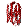
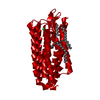
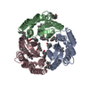
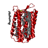
Density Matthews: 2.7 Å3/Da / Density % sol: 54.48 % Description: ISOMORPHOUS STRUCTURE OF CHLORIDE-BOUND HALORHODOPSIN, PDB ENTRY 5AHY
Crystal grow
Temperature: 296 K / Method: vapor diffusion Details: RESERVOIR SOLUTION: 100 MM BICINE PH 8, 150 MM NACL, 2.4 M (NH4)2SO4. VAPOR DIFFUSION AT 296 K WITH DROP RATIO OF 1.2 TO 0.8 UL PROTEIN TO RESERVOIR. PROTEIN CONCENTRATION: 4.9 MG/ML. POST- ...Details: RESERVOIR SOLUTION: 100 MM BICINE PH 8, 150 MM NACL, 2.4 M (NH4)2SO4. VAPOR DIFFUSION AT 296 K WITH DROP RATIO OF 1.2 TO 0.8 UL PROTEIN TO RESERVOIR. PROTEIN CONCENTRATION: 4.9 MG/ML. POST-CRYSTALLIZATION SOAKING SOLUTION: 100 MM GLYCINE PH 10, 2.6 M (NH4)2SO4 PH 10, 25% SUCROSE.
Monochromator: CRYSTAL SI(111) / Protocol: SINGLE WAVELENGTH / Monochromatic (M) / Laue (L): M / Scattering type: x-ray
Radiation wavelength
Wavelength: 0.9202 Å / Relative weight: 1
Reflection
Resolution: 2.6→45.5 Å / Num. obs: 8774 / % possible obs: 99.9 % / Observed criterion σ(I): -3 / Redundancy: 8.2 % / Biso Wilson estimate: 31.77 Å2 / Rmerge(I) obs: 0.16 / Net I/σ(I): 14.5
Reflection shell
Resolution: 2.6→2.73 Å / Redundancy: 5.7 % / Rmerge(I) obs: 0.91 / Mean I/σ(I) obs: 1.8 / % possible all: 99.8
-
Processing
Software
Name
Version
Classification
PHENIX
(PHENIX.REFINE)
refinement
XDS
datareduction
XSCALE
datascaling
PHENIX
phasing
Refinement
Method to determine structure: OTHER Starting model: NONE Resolution: 2.6→42.393 Å / σ(F): 1.34 / Stereochemistry target values: ML Details: THE WHOLE SECTION C-TERMINAL TO ALA261 IS DISORDERED
Rfactor
Num. reflection
% reflection
Rfree
0.2527
434
4.9 %
Rwork
0.1912
-
-
obs
0.1943
8763
99.8 %
Solvent computation
Shrinkage radii: 0.9 Å / VDW probe radii: 1.11 Å / Solvent model: FLAT BULK SOLVENT MODEL
Displacement parameters
Biso mean: 36.1 Å2
Refinement step
Cycle: LAST / Resolution: 2.6→42.393 Å
Protein
Nucleic acid
Ligand
Solvent
Total
Num. atoms
1822
0
41
13
1876
Refine LS restraints
Refine-ID
Type
Dev ideal
Number
X-RAY DIFFRACTION
f_bond_d
0.008
1930
X-RAY DIFFRACTION
f_angle_d
0.999
2648
X-RAY DIFFRACTION
f_dihedral_angle_d
17.574
1099
X-RAY DIFFRACTION
f_chiral_restr
0.052
329
X-RAY DIFFRACTION
f_plane_restr
0.006
316
LS refinement shell
Resolution (Å)
Rfactor Rfree
Num. reflection Rfree
Rfactor Rwork
Num. reflection Rwork
Refine-ID
% reflection obs (%)
2.6001-2.9763
0.2791
149
0.1992
2721
X-RAY DIFFRACTION
100
2.9763-3.7494
0.2672
141
0.1885
2762
X-RAY DIFFRACTION
100
3.7494-42.3988
0.2343
144
0.1895
2846
X-RAY DIFFRACTION
100
+
About Yorodumi
-
News
-
Feb 9, 2022. New format data for meta-information of EMDB entries
New format data for meta-information of EMDB entries
Version 3 of the EMDB header file is now the official format.
The previous official version 1.9 will be removed from the archive.
In the structure databanks used in Yorodumi, some data are registered as the other names, "COVID-19 virus" and "2019-nCoV". Here are the details of the virus and the list of structure data.
Jan 31, 2019. EMDB accession codes are about to change! (news from PDBe EMDB page)
EMDB accession codes are about to change! (news from PDBe EMDB page)
The allocation of 4 digits for EMDB accession codes will soon come to an end. Whilst these codes will remain in use, new EMDB accession codes will include an additional digit and will expand incrementally as the available range of codes is exhausted. The current 4-digit format prefixed with “EMD-” (i.e. EMD-XXXX) will advance to a 5-digit format (i.e. EMD-XXXXX), and so on. It is currently estimated that the 4-digit codes will be depleted around Spring 2019, at which point the 5-digit format will come into force.
The EM Navigator/Yorodumi systems omit the EMD- prefix.
Related info.:Q: What is EMD? / ID/Accession-code notation in Yorodumi/EM Navigator
Yorodumi is a browser for structure data from EMDB, PDB, SASBDB, etc.
This page is also the successor to EM Navigator detail page, and also detail information page/front-end page for Omokage search.
The word "yorodu" (or yorozu) is an old Japanese word meaning "ten thousand". "mi" (miru) is to see.
Related info.:EMDB / PDB / SASBDB / Comparison of 3 databanks / Yorodumi Search / Aug 31, 2016. New EM Navigator & Yorodumi / Yorodumi Papers / Jmol/JSmol / Function and homology information / Changes in new EM Navigator and Yorodumi
 Movie
Movie Controller

 Yorodumi
 Open data
 Basic information
 Components
 Keywords
 Function and homology information
 Authors
 Citation
 Structure visualization
 Downloads & links
 Other downloads
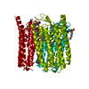
 PDBj
PDBj


 Assembly

 Mass: 284.436 Da / Num. of mol.: 1 / Source method: obtained synthetically / Formula: C20H28O
 Mass: 35.453 Da / Num. of mol.: 1 / Source method: obtained synthetically / Formula: Cl
 Type: D-saccharide / Mass: 292.369 Da / Num. of mol.: 1
 Mass: 18.015 Da / Num. of mol.: 13 / Source method: isolated from a natural source / Formula: H2O
 Sample preparation
 / Beamline: X10SA / Wavelength: 0.9202
  Processing