- PDB-2jag: L1-intermediate of halorhodopsin T203V -
+
Open data
ID or keywords:
Loading...
-
Basic information
Entry
Database: PDB / ID: 2jag
Title
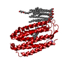
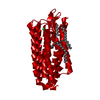
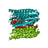
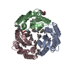
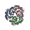
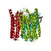
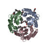
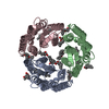
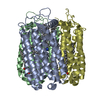
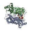
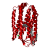
L1-intermediate of halorhodopsin T203V
Components
Halorhodopsin
Keywords
MEMBRANE PROTEIN / CHROMOPHORE / CHLORIDE PUMP / ION TRANSPORT / SENSORY TRANSDUCTION / MEMBRANE / CHLORIDE / RECEPTOR / ION PUMP / TRANSPORT / PHOTORECEPTOR PROTEIN / TRANSMEMBRANE / RETINAL PROTEIN / INTERMEDIATE L1
Function / homology
Function and homology information
photoreceptor activity / phototransduction / monoatomic ion channel activity / plasma membrane Similarity search - Function
Resolution: 1.93→20 Å / Data cutoff high absF: 10000 / Cross valid method: THROUGHOUT / σ(F): 0 Details: 3-10 HELIX DISTORTION BETWEEN LEU A 110 AND ALA A 113. PI-BULGE BETWEEN ALA A 178 AND TRP A 183 DISRUPTS HELIX E.RETINAL IN 13-CIS CONFORMATION.
Rfactor
Num. reflection
% reflection
Selection details
Rfree
0.2608
959
4.6 %
RANDOM
Rwork
0.2373
-
-
-
obs
0.2373
19178
91.9 %
-
Solvent computation
Bsol: 99.7591 Å2 / ksol: 0.488806 e/Å3
Displacement parameters
Baniso -1
Baniso -2
Baniso -3
1-
0.859 Å2
0.219 Å2
0 Å2
2-
-
0.859 Å2
0 Å2
3-
-
-
-1.718 Å2
Refinement step
Cycle: LAST / Resolution: 1.93→20 Å
Protein
Nucleic acid
Ligand
Solvent
Total
Num. atoms
1822
0
60
41
1923
Refine LS restraints
Refine-ID
Type
Dev ideal
X-RAY DIFFRACTION
c_bond_d
0.015
X-RAY DIFFRACTION
c_bond_d_na
X-RAY DIFFRACTION
c_bond_d_prot
X-RAY DIFFRACTION
c_angle_d
X-RAY DIFFRACTION
c_angle_d_na
X-RAY DIFFRACTION
c_angle_d_prot
X-RAY DIFFRACTION
c_angle_deg
1.4
X-RAY DIFFRACTION
c_angle_deg_na
X-RAY DIFFRACTION
c_angle_deg_prot
X-RAY DIFFRACTION
c_dihedral_angle_d
X-RAY DIFFRACTION
c_dihedral_angle_d_na
X-RAY DIFFRACTION
c_dihedral_angle_d_prot
X-RAY DIFFRACTION
c_improper_angle_d
X-RAY DIFFRACTION
c_improper_angle_d_na
X-RAY DIFFRACTION
c_improper_angle_d_prot
X-RAY DIFFRACTION
c_mcbond_it
X-RAY DIFFRACTION
c_mcangle_it
X-RAY DIFFRACTION
c_scbond_it
X-RAY DIFFRACTION
c_scangle_it
+
About Yorodumi
-
News
-
Feb 9, 2022. New format data for meta-information of EMDB entries
New format data for meta-information of EMDB entries
Version 3 of the EMDB header file is now the official format.
The previous official version 1.9 will be removed from the archive.
In the structure databanks used in Yorodumi, some data are registered as the other names, "COVID-19 virus" and "2019-nCoV". Here are the details of the virus and the list of structure data.
Jan 31, 2019. EMDB accession codes are about to change! (news from PDBe EMDB page)
EMDB accession codes are about to change! (news from PDBe EMDB page)
The allocation of 4 digits for EMDB accession codes will soon come to an end. Whilst these codes will remain in use, new EMDB accession codes will include an additional digit and will expand incrementally as the available range of codes is exhausted. The current 4-digit format prefixed with “EMD-” (i.e. EMD-XXXX) will advance to a 5-digit format (i.e. EMD-XXXXX), and so on. It is currently estimated that the 4-digit codes will be depleted around Spring 2019, at which point the 5-digit format will come into force.
The EM Navigator/Yorodumi systems omit the EMD- prefix.
Related info.:Q: What is EMD? / ID/Accession-code notation in Yorodumi/EM Navigator
Yorodumi is a browser for structure data from EMDB, PDB, SASBDB, etc.
This page is also the successor to EM Navigator detail page, and also detail information page/front-end page for Omokage search.
The word "yorodu" (or yorozu) is an old Japanese word meaning "ten thousand". "mi" (miru) is to see.
Related info.:EMDB / PDB / SASBDB / Comparison of 3 databanks / Yorodumi Search / Aug 31, 2016. New EM Navigator & Yorodumi / Yorodumi Papers / Jmol/JSmol / Function and homology information / Changes in new EM Navigator and Yorodumi
 Movie
Movie Controller


 Open data
 Basic information
 Components
 Keywords
 Function and homology information
 Authors
 Citation
 Structure visualization
 Downloads & links
 Other downloads

 PDBj
PDBj


 Assembly

 Type: D-saccharide / Mass: 292.369 Da / Num. of mol.: 1


 Mass: 35.453 Da / Num. of mol.: 2 / Source method: obtained synthetically / Formula: Cl
 Mass: 256.424 Da / Num. of mol.: 1 / Source method: obtained synthetically / Formula: C16H32O2
 Mass: 284.436 Da / Num. of mol.: 1 / Source method: obtained synthetically / Formula: C20H28O
 Sample preparation
 / Beamline: ID14-2 / Wavelength: 0.9393
  Processing